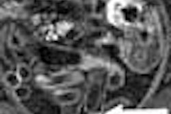
There are a number of reasons why multidetector-row CT is making inroads in rectal cancer staging. Perhaps none is more compelling, though, than the ability to look for metastases and stage the cancer based on a single CT scan.
"We're already doing CT on rectal cancer patients; they come to us for abdomen/pelvis CTs to look for metastatic disease," said Dr. Terry Desser, an associate professor of radiology at Stanford University in Stanford, CA. With the aid of a protocol her group has modified slightly, rescanning to stage the cancer is often unnecessary, she said.
Speaking at the 2004 International Symposium on Multidetector-row CT in San Francisco, Desser discussed the use of CT for rectal cancer staging at her institution, comparing it to MRI and the current gold standard, endorectal ultrasound.
Colorectal cancer is the third most common cause of cancer in the U.S., with 150,000 new U.S. cases expected in 2004, according to American Cancer Society statistics. But those cancers aren't evenly distributed. About 42,000 of the new cases will be located in the rectum, with another quarter of the lesions in the sigmoid colon, and the rest distributed fairly evenly in the colon, Desser said.
"Treatment strategies are based on achieving local and regional control over the disease," Desser said. "Treatment varies greatly by stage." As a result, she said, locoregional control of the disease through surgery and radiotherapy is crucial for maintaining good outcomes, and to enable sphincter-sparing surgery instead of more radical options whenever possible.
Patients with locally advanced disease get preoperative chemotherapy and/or radiation before surgery, while those with less advanced disease go straight to surgery. "So if we can determine the subgroup of patients with locally advanced disease, we can help to direct therapy," she said.
Tumor classifications
Staging systems are used to designate the depth and invasion of the primary tumor, and assess the depth and extent of the spread, she said. The Dukes' classifications of old (A, B, and C) have, of course, been supplanted by the American Joint Committee on Cancer's (AJCC) TNM classification system, which looks separately at the extent of tumors, nodes, and metastases.
"T" refers to the depth of tumor invasion. T1 tumors are confined to the mucosa or submucosa, T2 tumors involve the muscle layers of the colon, while T3 tumors invade the muscularis propria and into the subserosal fat or perirectal adipose tissue, Desser said. Stage T4 represents tumors that have invaded surrounding organs, such as the prostate, uterus, or blood vessels.
The just-released sixth edition of the AJCC staging system refines this process even further for T staging by stratifying stages II and III into IIa, IIb, IIIa, IIIb, and IIIc. The newest classification also includes the number of positive lymph nodes detected (N) compared to a simpler N-staging scheme in the previous version consisting of N (no positive nodes), N1 (1-3 positive nodes), or N2 (3 or more positive nodes). The M stage (0 or 1), indicating the presence or absence of metastases, is unchanged in version 6.
Imaging techniques
MRI can provide "spectacular images," but is more expensive and cumbersome than CT, requiring two acquisitions to provide what CT can do in a single exam, Desser said. And while the current gold standard, endorectal ultrasound (EUS), can also provide good results, at least for T and N staging, its accuracy suffers from widely varying sensitivity at different disease stages.
A 1992 textbook by a leading practitioner reported that EUS' T-stage accuracy ranged from 94% for T1 disease, to 72% for stage T2 cancer, to 92% for stage T3 and 94% for T4. Accuracy for N-stage disease ranged from 16% to 86%, and its sensitivity for detecting transmural tumor infiltration ranged from 50% to 90%, Desser said (Gastroenterologic Endosonography by T. Rosch).
"One of most important limitations is that when you have advanced rectal cancer, it becomes impossible to pass the (EUS) endoscope up across the narrow lumen that straddles the lesions," Desser said. "Many of the studies of endorectal ultrasound have had to exclude up to almost a quarter of the cases. For very high rectal lesions, you have the problem of poor acoustic coupling."
Microtumoral and peritumoral infiltration, as well as fibrosis, present additional problems. And of course, EUS offers only limited visualization of lymph nodes and other surrounding tissues.
Single-slice CT has proved to be an ideal replacement. However, MDCT, with its thin-section imaging and the availability of multiplanar reconstructions (MPR), may offer more accurate local staging than EUS, Desser said.
"MPRs are really the big selling points to this technique, because they really show the surgeon something they can't see with endoluminal ultrasound," she said.
Surgical goals
Locoregional control is crucial for preventing disease recurrence, and the surgical technique chosen can also have a great impact on outcomes, Desser said. Nowadays, surgeons endeavor to spare the anal sphincter whenever possible to avoid the need for a permanent colostomy. They manage to do so in nearly three-fourths of rectal cancer cases.
The sphincter muscles can often be spared for good surgical candidates with least 2 cm clearance between the tumor and the anal verge -- though the distance from the anal verge is generally determined by rigid sigmoidoscopy, not imaging, Desser said.
The superior rectal artery provides the principal blood supply for the rectum, with varying contributions from the middle rectal and inferior rectal arteries. "The thing about the arterial supply is that it's the landmark for lymphatic drainage of the rectum," she said. As rectal cancer progresses, "we'll typically see nodal spread first into the perirectal flap and up along the superior rectal artery and into the IMA (inferior mesenteric artery)."
Acquisition
Generally the rectal cancer is confirmed endoscopically, then the patients come to imaging for staging, Desser said. In order to combine staging and the search for metastatic disease, her group has developed a modified scan technique consisting of a standard abdomen/pelvis scan, but with thinner sections through the pelvis.
After cleansing with two Fleet enemas, extension of the rectal lumen before imaging is typically performed with warm water rather than air, she said. This provides better definition of the rectal wall architecture, prevents spiral artifacts, and is somewhat easier to maintain than air.
Contrast administration (300-350 mgL/mL iodine at 40 sec at 4 cc/sec) begins at the same time as the water distension. "The arterial phase is useful for seeing the blush of early (lesion) enhancement," she said. "Venous phase is good for avoiding confusing small veins and lymph nodes."
Following a 70-second scan delay, images of the abdomen and pelvis are acquired on an 8- or 16-slice MDCT scanner, using 8 x 1.25 or 16 x 1.25 detector configuration to enable retrospective reconstructions. Slices of 5-mm thickness are obtained from the diaphragm to the iliac crest, with 2.5-mm slices from the crest to 3 cm below the pubis. Pelvic images are reconstructed at 1.25-mm slice thickness and 0.6-mm interval, using a 20-cm field-of-view centered on the rectum.
Interpretation
Desser's group creates various images on the workstation, including curved planar sagittal and coronal images and oblique axial images through the true rectal short axis.
"The key to CT interpretation is of course to find the lesion, which often enhances, or we'll just see an intraluminal mass that contrasts with the water we've got in there," she said. "We evaluate the contour of the outer rectal wall, (and) look closely at its interface with fat to see if there is any nodularity or spiculation. We look at the boundary with adjacent organs, like the prostate and uterus. We look at the endopelvic fascia to see whether it's thickened on one side, or whether there is extension of tumor up to it or nodes external to it, and then we look for lymph nodes or distal metastases."
The lymph nodes follow the arterial supply, Desser said. Her group uses a 5-mm size cutoff for reporting nodes. The valves of Houston can sometimes simulate a lesion. And while size criteria are often unreliable for nodal staging, irregular borders can be indicative of involvement, she said.
"You can make some nice volume-rendered images, but fundamentally what you need to know is on the axial images," Dresser said.
"Our feeling is that MDCT can provide some very useful information about the local extent of rectal cancers, as well as distal metastatic disease, and that it's useful in surgical and radiation therapy planning," she said.
By Eric Barnes
AuntMinnie.com staff writer
October 25, 2004
Related Reading
Rectal cancer chemo, radiation best before surgery, October 21, 2004
Revised colon cancer staging provides better survival estimates, October 13, 2004
Combined PET/CT accurately detects pelvic recurrence of rectal cancer, September 8, 2004
Preoperative approach may benefit patients with locally advanced rectal cancer, February 6, 2004
PET/CT boosts confidence in rectal cancer staging, US yields high sensitivity, April 3, 2003
Copyright © 2004 AuntMinnie.com


