A new study from Hong Kong suggests that whole-body PET/CT scans and excessive radiation doses can increase the risk of cancer, especially among young patients and women.
The research from the University of Hong Kong and Queen Mary Hospital in Hong Kong also offers effective CT radiation doses for men and women, as well as effective doses for combined PET/CT procedures.
PET/CT "has a very important role in oncology and infection, and its role has been expanding," said Dr. Bingsheng Huang from the University of Hong Kong's Department of Diagnostic Radiology and Department of Clinical Oncology. "However, it has increased patient radiation exposure and this radiation has a well-known, adverse effect on patients" with a greater cancer risk.
The purpose of the study, presented at the 2008 American Roentgen Ray Society (ARRS) meeting in Washington, DC, was to estimate the radiation dose of an adult whole-body PET/CT with a diagnostic CT scan and clinically evaluate the cancer risk to the Hong Kong population caused by the radiation exposure.
Researchers analyzed whole-body FDG PET/CT scans from vertex to midthigh acquired on a Discovery PET/CT system from GE Healthcare of Chalfont St. Giles, U.K. The hybrid scanner is equipped with 64-slice CT technology, and the injected dose of F-18 FDG was assumed to be 370 MBq. Direct measurements of CT scan dose were performed on a standard phantom equipped with 314 thermoluminescent dosimeters.
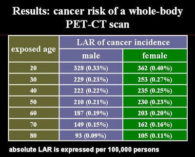
The lifetime attributable risk (LAR) of cancer incidence was used to estimate the excessive risk caused by the radiation of a PET/CT scan. The study's source for that data was the "Health Risks from Exposure to Low Levels of Ionizing Radiation: BEIR VII Phase 2" report, published by the National Academy of Sciences in Washington, DC, in 2006. Researchers used the information to create a model to calculate the lifetime attributable risk for the Hong Kong Chinese population.
"In this (BEIR) report, each organ's cancer risk is given and each organ cancer risk is calculated for a special radiation dose," Huang said in his presentation. "The whole-body cancer risk can be acquired by summing up each organ's risk."
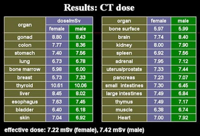
The researchers found that the effective radiation dose for a CT scan is 7.22 mSv for women and 7.42 mSv for men. The effective dose for a PET scan was calculated to be 6.23 mSv for both women and men. Huang noted a high radiation PET dose of 59.20 mSv in the bladder, attributing the finding to the accumulation of FDG in the bladder.

Subsequently, the total combined effective radiation dose for PET/CT is 13.45 mSv for women and 13.65 mSv for men.
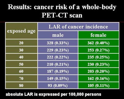
Using the LAR model, the researchers also calculated the cancer risk for men and women between the ages of 20 and 80 years. "For males, the cancer risk is from 0.33% to 0.09% (per 100,000 men). For females, it is from 0.40% to 0.11% (per 100,000 women)," Huang said. "We can see that the cancer risk increased when the exposed age decreased, and the females' cancer risk is always higher than males'."

The researchers calculated that the cancer incidence induced by one whole-body PET/CT scan contributes approximately 1% to 2% to total lifetime cancer incidence. Given this finding, Huang said that the radiology community should be mindful of the situation and clinically justify the use of PET/CT on patients.
The researchers further recommended that "modification of scanning protocols tailored to the specific clinical indication should be considered, so as to balance between good image quality and reduced radiation dose exposure."
By Wayne Forrest
AuntMinnie.com staff writer
May 5, 2008
Related Reading
Researchers recommend alternatives to CT, April 15, 2008
ACC news: CTA dose varies widely, could be lower, April 2, 2008
Prospective gating drops cardiac CT radiation dose, March 10, 2008
New studies examine CR, CT radiation dose, March 9, 2008
ACR responds to NEJM article on CT radiation risks, November 29, 2007
Copyright © 2008 AuntMinnie.com


