In an era of healthcare cuts that often lead to fewer procedures being performed, existing CT data offer the rare advantage of a free extra test -- namely a bone mineral density study that's there for the taking but that usually goes unused, according to an article in the Annals of Internal Medicine.
In a retrospective study of more than 2,000 patients, researchers from the University of Wisconsin in Madison found that measuring CT attenuation in the vertebrae (specifically L1) correctly classified more than 80% of patients as positive or negative for osteoporosis.
CT results hewed closely to those obtained with the gold standard, dual-energy x-ray absorptiometry (DEXA). The comparisons were accurate at multiple levels of osteoporosis severity, though CT is actually a superior test that doesn't confuse compression fractures of the spine with healthier bone density levels, the authors wrote (Ann Intern Med, April 15, 2013).
"This is free information that's easy to get at, and it's being underused right now," said lead author Dr. Perry Pickhardt in an interview with AuntMinnie.com. "The CT is being done for another reason, so we're basically just using the information without any additional dose, cost, or time to the patient, and it can not only be done as we read these, we can go back in time and pull the study off the PACS, and that's what we did."
The U.S. Preventive Services Task Force recommends bone mineral density (BMD) testing with DEXA in women 65 years and older, or in younger individuals with risk levels at least as high. But more than half of female Medicare beneficiaries have never been screened with DEXA, and more than 80% of patients with an osteoporosis fracture have never been tested or received drug treatment. On the other hand, the tens of millions of CT scans of the chest or abdomen acquired each year could readily be used for osteoporosis screening, the authors stated.
The study evaluated bone mineral density from existing CT scans compared with existing DEXA data in a smaller patient population. The study focused on the L1 vertebrae because it is easily identifiable as the first nonrib-bearing vertebra on thoracic and abdominal scans, Pickhardt said, adding that levels L1 through L5 all were evaluated and showed little variation.
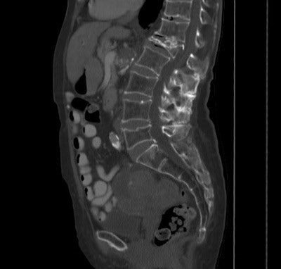 Above and below: In a patient with an L1 compression fracture, low attenuation in the L2 vertebra (27 HU) was used to diagnose osteoporosis at CT. All images courtesy of Dr. Perry Pickhardt.
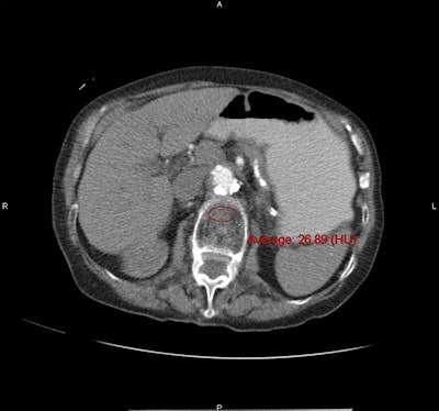
Along with the conventional DEXA exams, CT bone mineral density was measured by choosing a region of interest over vertebral trabecular bone, and measuring Hounsfield units, being careful not to exclude areas that might distort the reading.
The CT exams had been performed for various reasons, including suspected masses or oncologic workup, gastrointestinal or genitourinary concerns, abdominal pain investigations, and virtual colonoscopy screening.
The assessment parameters for CT could be set to detect osteoporosis with either 90% sensitivity or 90% specificity, depending on the goals of the test. Sensitivity, specificity, and positive and negative predictive values were based on kernel-density plots of L1 CT attenuation for normal, osteopenic, and osteoporotic groups, the authors noted.
Thus, DEXA results applied across the range of CT attenuation results were used to set thresholds that would yield either the 90% sensitivity or 90% sensitivity outcome, or a balance between the two when needed to distinguish osteoporosis from osteopenia. Specifically, an attenuation threshold of 160 HU or less was 90% sensitive, and a threshold of 110 HU was more than 90% specific for distinguishing osteoporosis from osteopenia and normal bone mineral density, the group reported.
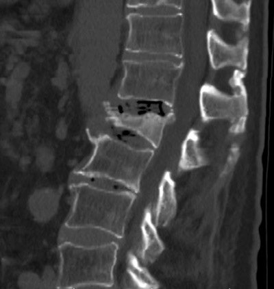 DEXA results were normal in a patient with an L1 compression fracture and osteoporosis at CT.
CT attenuation values were significantly lower at all vertebral levels for patients with osteoporosis at DEXA (p < 0.001) at the L1 level. Positive predictive values for osteoporosis were 68% or greater at CT attenuation thresholds less than 100 HU; negative predictive values were 99% at thresholds greater than 200 HU, the group reported.
Almost 23% of the patients had osteoporosis, 45% had intermediate bone mineral density or osteopenia, and 32% had normal bone density.
"You can't really talk in terms of accuracy, because it's very hard to compare the DEXA [to CT], but it's pretty clear that when somebody has a 100 HU or lower in the spine, [the patient] is very likely to have osteoporosis, and they might even have a fracture that we can directly see," Pickhardt said. "A lot of these people have been unscreened, or even missed at DEXA, so it's something that radiologists and others need to be aware of."
The method hasn't been validated in terms of establishing agreed-upon HU thresholds, but the study data are a good step in that direction that others can build on by measuring CT-based bone mineral density in their patients who have received CT of the abdomen for another reason, Pickhardt said.
Questions incidental findings
In an editorial accompanying the article, Dr. Sumit R. Majumdar from the University of Alberta and Dr. William Leslie from the University of Manitoba in Canada argued that at this stage of investigation the higher specificity threshold should be favored even if it reduced sensitivity, in order to minimize the possibility of false-positive results.
Rather than ruling out osteoporosis, Majumdar and Leslie believe the CT exam would best be used to rule in osteoporosis identified in DEXA scans, they wrote. For one thing, medical records are already replete with findings that are never acted on, and second, "the physician left to deal with osteoporosis will be neither the specialist who ordered the CT scan nor the reporting radiologist, [so] we should have low tolerance for false-positive results," the team wrote.
But Pickhardt found that suggestion to be unnecessarily conservative, considering that patients can be considered individually, an issue that was covered in the article, Pickhardt said.
"If you have a high-risk population, I think going for high sensitivity makes more sense, because even if they turn out to be false-positive and they get a DEXA that shows osteopenia or normal [density], I would actually believe the CT," he said. "But if it's a low-risk population, let's say a 45-year-old guy who has a very low apriori risk of osteoporosis, you don't want to use a high-sensitivity threshold, you want to use a high-specificity threshold because we don't want a false positive."
The authors of the editorial also cited the lack of CT hip data in the analysis, since hips are the site of so many debilitating fractures. But Pickhardt said there are good data showing that whether the vertebrae are normal or at risk, the hips follow suit. And a current project addresses this issue by using software cleared by the U.S. Food and Drug Administration to create DEXA-equivalent T-scores from hip data, turning it into something like a planar measurement, Pickhardt said.
"The good news there is that it directly correlates to DEXA," which is probably a little more reliable in the hip than in the spine, he said. Imperfect or not, the CT results are easy to obtain, and the data are widely available in the population at risk for osteoporosis, he said.
"If your Aunt Edna had a CT scan recently, I can just pull up her scan and tell you if her bone density is normal, abnormal, or whatever," Pickhardt said. "It's a win for CT, and it's another way to add value in this era of cost-consciousness."


