A radiologist from Alabama who was one of the first healthcare professionals to receive the COVID-19 vaccine last week described her experience in a December 18 article in the Business Insider.
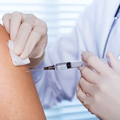
Dr. Joy Henningsen works as a diagnostic radiologist at Birmingham Veterans Affairs Medical Center. She received the Pfizer/BioNTech vaccine on December 17 along with other personnel at the facility. The Birmingham VA Medical Center received some of the first shipments of the vaccine in the U.S.
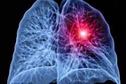
Henningsen decided to share her story in a first-person article she wrote for Business Insider because of her experiences interpreting chest and lung images of patients with COVID-19. She noted that while as a radiologist she's not on the front lines in caring for COVID-19 patients, she sees the damage caused by the SARS-CoV-2 virus through their images.
"I am a constant witness to COVID-19's serious effects via the dark tales told by my patients' medical images," she wrote.
Henningsen also noted that she's an "outspoken pro-mask, pro-vaccine healthcare communicator" who felt compelled to tell her story to assuage concerns that some individuals may have about the vaccine's efficacy or its side effects. She even shared her enthusiasm via a post on her Twitter account.
My thoughts on the vaccine 😂 Who’s with me? #MedTwitter pic.twitter.com/Qb9STy3Qlp
— Joy Henningsen, MD 😷🗽 (@JoyHenningsenMD) December 2, 2020
Henningsen said getting the COVID-19 vaccine was "much less painful" than the flu shot she received earlier in the fall, and she's looking forward to receiving the second shot in three weeks.
Despite being vaccinated, she said she does not plan to return to a normal life of traveling and socializing until 75% of the American population is immunized.