A deep-learning (DL) model quantifies coronary plaque volume from coronary CT angiography (CCTA) images -- with strong correlation to radiologist interpretation and intravascular ultrasound (IVUS), researchers have reported.
"[The model] provided fully automated measurements of plaque volume from CCTA that closely agreed with expert readers and IVUS and carried prognostic value for future MACEs [major adverse cardiac events]," wrote a team led by Qian Chen, MD, of Nanjing Medical University in China. The group's findings were published April 21 in Radiology.
CCTA has been established as a first-line noninvasive technique for the assessment of coronary artery disease and can be used not only to evaluate coronary stenosis, but also to quantify plaque volume of the "whole-coronary artery tree atherosclerosis," Chen and colleagues explained. AI -- specifically DL algorithms -- can increase the efficiency and accuracy of CCTA image analysis, but the "prognostic value [of DL models] in monitoring plaque progression remains unknown," they wrote.
The team sought to develop and test a DL model it named PlaqueSegNet for quantifying plaque on CCTA images via a study that included 2,013 patients who underwent CCTA were retrospectively enrolled from 17 Chinese hospitals between June 2009 and May 2024. Imaging data from these exams were split into training (n=1,409) and validation sets (n=604) to develop the model. The group externally tested PlaqueSegNet using four independent datasets: a paired CCTA and intravascular US (IVUS) dataset; a subset of the CT-FFR-CHINA (study 3) dataset collected with different CT scanners; a serial CCTA dataset of exams taken within a three-month interval; and a photon-counting CT (PCCT) dataset.
Chen and colleagues evaluated PlaqueSegNet's prognostic value using the Harrell C-index in three cohorts:
- CT-FFR-CHINA study 2 (median follow-up, 2.3 years),
- CT-FFR-CHINA study 1.1 (median follow-up, 5.3 years), and
- A serial CCTA cohort (median follow-up, 3.6 years).
Overall, the group reported that PlaqueSegNet showed high agreement and reproducibility for quantifying plaque volume against IVUS and expert readers across the four external datasets (all intraclass correlation coefficients, >0.90). It also found that the model's predictive performance held up over longer follow-up:
Performance of PlaqueSegNet for predicting major adverse cardiac events (MACEs) across various study cohorts | |
Study cohort | C-index |
| CT-FFR-CHINA study 2 | 0.64 |
| CT-FFR-CHINA study 1.1 | 0.65 |
| Serial CCTA | 0.74 |
The researchers also reported that the model cut analysis time for expert readers from roughly 19 minutes to under 11 seconds per patient.
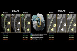
 Two representative cases demonstrate the performance of PlaqueSegNet for coronary plaque segmentation. (A–D) Coronary CT angiography (CCTA) and intravascular US (IVUS) images in a 65-year-old male patient with chest pain for two months show plaque. (A) Cross-sectional IVUS image shows a lipid-rich plaque (hypoechoic, arrows). (B) Axial and (C) cross-sectional CCTA images show the same plaque (low-attenuation area, yellow line and arrows) in the proximal left anterior descending artery. (D) PlaqueSegNet segmentation of the noncalcified plaque (yellow shading) and coronary lumen (green shading) in the cross-sectional image. (E–G) CCTA images in a 57-year old female patient with chest pain for three months show multiple plaques. (E) Volume-rendered CCTA image of the coronary tree shows PlaqueSegNet plaque segmentation (yellow) in the right and left coronary arteries. (F, G) PlaqueSegNet segmentation delineates a mixed plaque (yellow shading) and coronary lumen (green shading) in the left main artery and proximal-to-mid left anterior descending artery.RSNA
They did acknowledge some study limitations, noting that Bland–Altman analysis showed wide ranges of agreement across all comparisons, meaning plaque volumes could be over- or underestimated by roughly 100 mm³ to 150 mm³ on standard CT, and that the training data -- taken from Chinese tertiary hospitals -- may limit the study findings' generalizability to other settings.
A prospective trial, Serial Coronary CTA-based Plaque Progression Detection for Management of Coronary Heart Disease (SUCCESS), is underway "to further validate PlaqueSegNet's predictive value in serial CT angiography," they wrote.
"The study demonstrates that an automated plaque quantification tool is feasible to develop, can approximate expert and intravascular US assessments, and contains prognostic information at scale," noted Michelle Williams, MBChB, PhD, of the University of Edinburgh in the U.K., in an accompanying editorial. But further research is needed.
"There remain reasons to be cautious, including imperfect agreement, dependence on image quality, selected training and testing data, and uncertain therapeutic implications," she concluded.
Access the full study here.


![Axial images from unenhanced calcium score cardiac CT (left) and curved planar reformation images from CT angiography (right) show that higher long-term exposure to air pollution is associated with greater coronary artery calcium and more obstructive coronary artery disease (CAD). Top row: Images in a 68-year-old male patient with higher 10-year mean ambient air pollution exposure (7.9 μg/m3 for particulate matter measuring ≤2.5 μm in diameter [PM2.5] and 17.4 parts per billion [ppb] for NO2) with extensive CAD (coronary artery calcium score [CACS] >1,000 and obstructive CAD [≥70% diameter stenosis]). Bottom row: Images in a 57-year-old female patient with lower 10-year mean ambient air pollution exposure (6.3 μg/m3 for PM2.5 and 4.6 ppb for NO2) with no CAD (CACS = 0 and no obstructive stenosis).](https://img.auntminnie.com/mindful/smg/workspaces/default/uploads/2026/06/hanneman.r6SMLzkezo.png?auto=format%2Ccompress&dpr=2&fit=crop&h=167&q=70&w=250)


