Up to 64% of patients treated with high-precision radiation therapy (HPRT) for lung cancer develop imaging findings that can mimic tumor recurrence, but only 10% of these are confirmed to be true disease relapse, according to a review published April 16 in RadioGraphics.
This statistic represents a diagnostic challenge that carries significant risk of unnecessary intervention, especially when it comes to radiation-induced lung injury (RILI), wrote a team led by Omar Andrés Pantoja-Burbano, MD, of Pontificia Universidad Javeriana-Hospital Universitario San Ignacio in Bogotá, Colombia.
"The appearance and temporal evolution of RILI associated with high-precision dose techniques are similar to those of conventional radiation therapy but have important differences," the group explained. "Knowledge of these differences is essential in the interpretation of imaging findings after HPRT and allows appropriate identification of local recurrence of malignancy and therapy-related complications."
HPRT techniques -- including stereotactic body radiation therapy, intensity-modulated radiation therapy, and proton therapy -- have significantly improved tumor targeting in lung cancer while minimizing damage to surrounding lung tissue. But despite these advances, RILI remains a common complication, often manifesting as acute pneumonitis and chronic fibrosis, the group explained. Compared to conventional radiation therapy, HPRT has been linked to delayed onset and more localized manifestations of RILI, and to make things more tricky, these manifestations can not only be delayed after treatment but can also mimic tumor recurrence, with mass-like or scar-like opacities.
Pantoja-Burbano and colleagues offered the following insights on how RILI can appear, noting that it progresses through two distinct phases:
- An acute pneumonitic phase tends to begin more than three months after HPRT completion -- later than with conventional radiotherapy -- and is marked by ground-glass opacities, consolidation, and sometimes, the reversed halo sign or the crazy paving pattern.
- A chronic fibrotic phase tends to emerge around nine months post-treatment and peaks between one and two years after treatment; it can continue to evolve for up to four years. Three patterns tend to manifest: a modified conventional atelectatic opacity (46% to 71% of cases); a mass-like opacity resembling recurrence (7% to 20%); and a linear, scar-like opacity (11% to 22%).
Distinguishing fibrosis from recurrence depends on recognizing high-risk CT features on serial imaging, the group explained, noting that bulging margins on nodules indicate 100% specificity for recurrence, while craniocaudal growth on nodules of 5 mm or more translates to 92% sensitivity. Opacity enlargement beyond 12 months reaches 100% sensitivity and 83% specificity, the team wrote, and noted that a combination of three or more high-risk CT features achieves over 90% sensitivity and specificity.
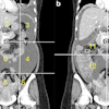
 Acute radiation-induced pneumonitis after SBRT (65 Gy, four fractions) in a 53-year-old woman with leiomyosarcoma of the uterus with a left lower lobe metastasis manifesting as a small nodule (not shown). (A) Axial dosimetric reconstruction of a CT image obtained for SBRT planning shows the metastasis receiving the maximal isodose (6500 cGy). (B) Axial CT image obtained 12 months after completion of RT shows focal ground-glass and consolidative opacities confined to the treatment plan (arrow). Note that acute lung injury with SBRT typically manifests later than with conventional RT (i.e., >12 weeks after completion of therapy), and in 25% of patients, the first CT manifestations occur more than one year after completion of RT.RadioGraphics
The bottom line? Although serial chest CT is the cornerstone of post-HPRT surveillance -- with major guidelines recommending imaging every three to six months for the first several years -- "accurate interpretation is critical to avoid misdiagnosis and unnecessary interventions," according to the group.
"Understanding the distinct radiologic features and timelines of RILI is essential for differentiating posttreatment changes from recurrent malignancy, guiding appropriate follow-up, and optimizing patient outcomes," it concluded.
Access the full article here.