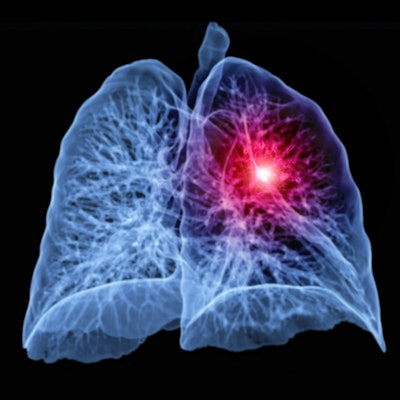
An artificial intelligence (AI) algorithm performs comparably to radiologists for reading low-dose CT (LDCT) lung cancer screening exams and could help reduce the workload in screening programs, according to a study published January 6 in Lung Cancer.
The ability to make CT lung cancer screening programs more efficient could be particularly helpful as demand ramps up due to expanded eligibility criteria, wrote a team led by Harriet Lancaster, PhD, of the University of Groningen in the Netherlands.
"Radiologists' challenging workloads associated with LDCT lung cancer screening is a noteworthy hurdle which needs to be addressed and overcome in the planning of lung cancer screening implementation," the group noted. "Artificial intelligence could offer a solution."
Studies have shown that lung cancer mortality can be reduced among high-risk populations through regular screening with low-dose chest CT. Although screening uptake has been lower than desired, recent changes to the eligibility pool may portend increased workload for radiologists. In March, the USPSTF lowered the starting age for lung cancer screening from 55 to 50 and reduced the number of pack-years from 30 to 20; in November, the U.S. Centers for Medicare and Medicaid Services followed suit.
With this in mind, the investigators evaluated AI's performance as a standalone reader of LDCT lung cancer baseline screening exams, comparing it with that of experienced radiologists. Their study included 283 people who had at least one solid lung nodule and who underwent low-dose CT between February 2017 and February 2018 as part of the Moscow Lung Cancer Screening initiative in Russia.
Under the study's protocol, five experienced thoracic radiologists assessed the nodules; the artificial intelligence lung cancer screening algorithm evaluated the nodules as well. Discrepancies between the radiologist readers, the AI algorithm, and a consensus reading of each exam (used as reference standard) were categorized either as positive or negative misclassifications.
There were 878 solid nodules found in the patient cohort. AI performed well compared with radiologist readers when classifying nodules that turned out to be negative and negative predictive value. However, AI misclassified a greater number of positive nodules, which the researchers attributed to overestimation of size of attached nodules -- a problem that could be addressed through further refinement of the AI system.
| AI vs. radiologists for classifying nodules on CT lung screening exams | ||
| Measure | Radiologist readers (range) | AI |
| Percentage of positive cases misclassified | 2.1%-8.8% | 18.7% |
| Percentage of negative cases misclassified | 1.4%-12.4% | 2.8% |
| Negative predictive value | 0.84-0.98 | 0.95 |
The team also calculated the extent to which AI could reduce radiologist workload in terms of the number of nodules that need to be worked up, finding a reduction that ranged from 77.4% to 86.7%.
Using AI as a standalone reader for lung cancer screening CT exams could be a very effective tool, the team concluded.
"To the best of our knowledge, we show for the first time that AI acting as an impartial reader in baseline screening can significantly reduce the radiologist's workload while not compromising on false-negative results of lung cancer screening with volume-based management of nodules and ultra-LDCT," it wrote. "Radiologists would then only need to read scans where nodules ≥ 100 mm3 are present in order to determine the follow-up strategy, instead of reading all scans."


