A deep-learning algorithm used with CT imaging can differentiate between benign and malignant vertebral fractures, researchers have found.
The algorithm proved comparable or better compared to radiologist readers, according to a team led by Sarah Foreman, MD, of Technische Universität München in Germany. The study findings were published March 26 in Radiology.
"[Deep-learning] models showed a high discriminatory power to differentiate between benign and malignant vertebral fractures, surpassing or matching the performance of radiology residents and matching that of a fellowship-trained radiologist," the group reported.
Distinguishing benign vertebral fractures from malignant ones can be tricky, and it's possible that using deep learning with CT could help, the team noted. The group conducted a study that investigated the reliability of CT-based deep learning and included 532 patients with benign or malignant vertebral fractures imaged between June 2005 and December 2022. The team developed three models, all of which used 3D U-Net encoder-classifier architecture.
The models' training set consisted of 381 patients (378 benign fractures, 447 malignant fractures, and 482 malignant lesions), while the internal test set included 86 patients and the external test set, 65 patients. The investigators assessed the models' performance using the area under the receiver operating characteristic curve (AUC) and compared results between two residents and one fellowship-trained radiologist.
The group found the following:
| AUC values from best performing deep-learning algorithm used with CT imaging to distinguish between benign and malignant vertebral fractures across a range of readers | ||
|---|---|---|
| Type of reader | Internal test set | External test set |
| All types | 0.91 | 0.76 |
| Resident reader 1 | 0.69 | 0.71 |
| Resident reader 2 | 0.7 | 0.71 |
| Fellowship-trained radiologist | 0.86 | 0.71 |
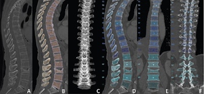
 Overview of the automated deep learning-based pipeline. Multidetector CT images show the (A) original data, (B, C) vertebral segmentation, and (D-F) vertebral labeling during preprocessing. Images and caption courtesy of the RSNA.
The study findings are useful, especially as "identifying the appropriate etiology of fractures is crucial to determine the most suitable therapeutic approach for patient treatment," wrote Christian Booz, MD, of the University Hospital Frankfurt, in Germany, and colleague Tommaso D'Angelo, MD, of University Hospital Messina in Italy in an accompanying commentary.
"[This study has] the potential to substantially improve fracture assessment and treatment in clinical routine," Booz and D'Angelo concluded.
The complete study can be found here.


