Contrast-enhanced CT improves the modality's ability to identify malignant lung nodules, researchers have found.
"[Our study demonstrated that a] logistic regression model based on plain and contrast-enhanced CT characteristics showed exceptional performance in the evaluation of malignancy for solitary solid lung nodules," wrote a team led by Wenjia Zhang, PhD, of Shanxi Medical University in China. "Utilizing this contrast-enhanced CT model [could] provide recommendations concerning follow-up or surgical intervention for preoperative patients presenting with [these] nodules." The research results were published September 19 in Nature: Scientific Reports.
Lung cancer is the primary cause of cancer death around the world, and imaging evaluation of solitary lung nodules is key to early detection and thus prompt and effective treatment, the group noted. Plain CT imaging is the go-to modality for identifying lung nodules, but its ability to differentiate benign from malignant lesions is limited. Techniques to characterize malignant nodules include tissue biopsy -- an invasive procedure -- and PET/CT, which can produce false negatives and positives and tends to be expensive. That's why adding contrast-enhanced CT shows promise for this task.
"In contrast to PET-CT, contrast-enhanced CT is relatively low-cost and remains the primary preoperative examination for most patients with lung nodules in developing countries," the group explained. "Contrast-enhanced CT helps to highlight blood vessels and other structures, making it easier to identify abnormalities such as tumors."
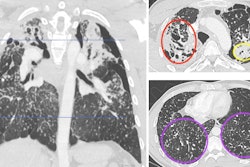
The group performed a study that compared the diagnostic performance of a plain CT-based model (model 1) and a plain CT plus a contrast-enhanced model (model 2) for classifying solitary solid pulmonary nodules as malignant. The research included 527 patients with lung nodules confirmed on pathology between January 2012 and July 2021. All patients underwent both plain and contrast-enhanced chest CT scans before undergoing surgery to remove the nodules.
Model 1 included two clinical characteristics and 15 plain CT characteristics, while model 2 included these plus four additional CT characteristics (enhanced CT value, enhancement rate, uniform enhancement, heterogeneous enhancement). The training cohort for the models consisted of 392 patients and the external validation cohort consisted of 135 patients; the authors assessed the diagnostic performance of each model using the area under the receiver operating curve (AUC) measure.
Study cohort patients presented preoperatively with the following:
- 201 malignant nodules (adenocarcinoma, 148 [73.6%]; squamous cell carcinoma, 35 [17.4%]; and large cell carcinoma,18 [9.0%]);
- 326 benign nodules (pulmonary hamartoma, 118 [36.2%]; sclerosing pneumocytoma, 35 [10.7%]; tuberculosis, 104 [31.9%]; and inflammatory pseudonodule, 69 [21.2%]).
The investigators found that the combination of plain CT plus contrast-enhanced CT improved performance measures for identifying malignant nodules.
| Comparison of two CT models for categorizing lung nodules as malignant (external validation cohort) | ||
|---|---|---|
| Measure (mean) | Model 1 (plain CT only) | Model 2 (plain CT plus contrast-enhanced CT) |
| AUC | 0.88 | 0.93 |
| Accuracy | 79% | 90% |
| Negative predictive value (NPV) | 87% | 93% |
| Positive predictive value (PPV) | 67% | 84% |
| Sensitivity | 79% | 88% |
| Specificity | 78% | 91% |
The bottom line? Model 2 "effectively reduces the missed diagnosis of lung cancer and avoids excessive surgery caused by misdiagnosis," the authors wrote.
"This … suggests that enhanced CT could be the basis for solitary solid pulmonary nodules preoperative diagnosis, especially when preoperative biopsy and PET-CT are not applicable," they concluded.
The complete study can be found here.