
Radiology vendors Fujifilm Healthcare and Qure.ai are partnering to combine their x-ray and artificial intelligence (AI) technologies to support tuberculosis (TB) screening in rural areas of Nigeria.
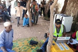
The companies are working with the Institute of Human Virology in Nigeria (IHVN) to deploy handheld x-ray systems with embedded AI technology as part of tuberculosis screening outreach efforts in the country. The project has the support of the U.S. Agency for International Development (USAID).
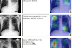
In the project, Fujifilm's FDR Xair portable handheld x-ray system will be used in combination with Qure.ai's qXR algorithms as part of rural mass testing programs to provide portable TB screening with high accuracy and efficiency at the point of care. Fujifilm and Qure.ai began working together to combine their technology in 2021.
The companies note that tuberculosis is the leading cause of death in sub-Saharan Africa; it is also the most opportunistic coinfection alongside HIV. The project also intends to detect drug-resistant tuberculosis and coinfections with HIV, and it covers contact tracing of presumptive TB cases to curb contagion and bring more cases into the national TB reporting system.


![Representative example of a 16-year-old male patient with underlying X-linked adrenoleukodystrophy. (A, B) Paired anteroposterior (AP) chest radiograph and dual-energy x-ray absorptiometry (DXA) report shows lumbar spine (L1 through L4) areal bone mineral density (BMD). The DXA report was reformatted for anonymization and improved readability. The patient had low BMD (Z score ≤ −2.0). (C) Model (chest radiography [CXR]–BMD) output shows the predicted raw BMD and Z score in comparison with the DXA reference standard, together with interpretability analyses using Shapley additive explanations (SHAP) and gradient-weighted class activation maps. The patient was classified as having low BMD, consistent with the reference standard. AM = age-matched, DEXA = dual-energy x-ray absorptiometry, RM2 = room 2, SNUH = Seoul National University Hospital, YA = young adult.](https://img.auntminnie.com/mindful/smg/workspaces/default/uploads/2026/04/ai-children-bone-density.0snnf2EJjr.jpg?auto=format%2Ccompress&fit=crop&h=100&q=70&w=100)


![Representative example of a 16-year-old male patient with underlying X-linked adrenoleukodystrophy. (A, B) Paired anteroposterior (AP) chest radiograph and dual-energy x-ray absorptiometry (DXA) report shows lumbar spine (L1 through L4) areal bone mineral density (BMD). The DXA report was reformatted for anonymization and improved readability. The patient had low BMD (Z score ≤ −2.0). (C) Model (chest radiography [CXR]–BMD) output shows the predicted raw BMD and Z score in comparison with the DXA reference standard, together with interpretability analyses using Shapley additive explanations (SHAP) and gradient-weighted class activation maps. The patient was classified as having low BMD, consistent with the reference standard. AM = age-matched, DEXA = dual-energy x-ray absorptiometry, RM2 = room 2, SNUH = Seoul National University Hospital, YA = young adult.](https://img.auntminnie.com/mindful/smg/workspaces/default/uploads/2026/04/ai-children-bone-density.0snnf2EJjr.jpg?auto=format%2Ccompress&fit=crop&h=112&q=70&w=112)


