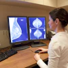
A study published March 24 in Radiology bears disturbing news: Radiologists are not able to easily distinguish AI-generated “deepfake” x-ray images from authentic ones.
In an interview with AuntMinnie, lead author Mickael Tordjman, MD, of the Icahn School of Medicine at Mount Sinai in New York, explained that the study involved 17 radiologists from 12 different centers in six countries. They were tested on two distinct image sets: first on a dataset of real and ChatGPT-4o-generated images of multiple anatomical regions and secondly on a dataset of half authentic chest x-rays and half created by RoentGen, an open-source generative AI model developed by Stanford Medicine researchers.
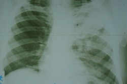
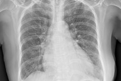
 Anatomy-matched real and GPT-4o-generated radiographs: (A) real and (B) GPT-4o-generated posteroanterior chest radiographs, (C) real and (D) GPT-4ogenerated lateral cervical spine radiographs, (E) real and (F) GPT-4o-generated posteroanterior hand radiographs, and (G) real and (H) GPT-4o-generated lateral lumbar spine radiographs. The pairs demonstrate that GPT-4o can produce radiographically plausible images across different anatomic regions. RSNA
What’s needed are tools to potentially help clinicians identify such deepfakes, Tordjman said. To that end, the study also identified common features of synthetic x-rays, and the authors have published a curated deepfake dataset with interactive quizzes for educational purposes.
"Deepfake medical images often look too perfect,” Tordjman said.
Potential solutions to clearly distinguish real and fake images and help prevent tampering include implementing advanced digital safeguards, such as invisible watermarks that embed ownership or identity data directly into the images and automatically attaching technologist-linked cryptographic signatures when the images are captured.
“We are potentially only seeing the tip of the iceberg,” Tordjman noted. “The logical next step in this evolution is AI-generation of synthetic 3D images, such as CT and MRI. Establishing educational datasets and detection tools now is critical.”
In an accompanying editorial, Rajesh Bhayana, MD, and Satheesh Krishna, MD, both of the University of Toronto, noted that the study demonstrates that reliance on visual inspection alone may no longer be sufficient for verifying image authenticity. The study is not an indictment of AI, however, but a call to augment imaging systems, they wrote.
“As the boundary between the authentic and the synthetic blurs, we must ensure that trust is built into the system itself. In the age of generative AI, seeing is no longer believing,” Bhayana and Krishna concluded.
The full study is available here.


![Representative example of a 16-year-old male patient with underlying X-linked adrenoleukodystrophy. (A, B) Paired anteroposterior (AP) chest radiograph and dual-energy x-ray absorptiometry (DXA) report shows lumbar spine (L1 through L4) areal bone mineral density (BMD). The DXA report was reformatted for anonymization and improved readability. The patient had low BMD (Z score ≤ −2.0). (C) Model (chest radiography [CXR]–BMD) output shows the predicted raw BMD and Z score in comparison with the DXA reference standard, together with interpretability analyses using Shapley additive explanations (SHAP) and gradient-weighted class activation maps. The patient was classified as having low BMD, consistent with the reference standard. AM = age-matched, DEXA = dual-energy x-ray absorptiometry, RM2 = room 2, SNUH = Seoul National University Hospital, YA = young adult.](https://img.auntminnie.com/mindful/smg/workspaces/default/uploads/2026/04/ai-children-bone-density.0snnf2EJjr.jpg?auto=format%2Ccompress&dpr=2&fit=crop&h=167&q=70&w=250)

