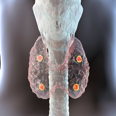
Choline-PET/CT should be the imaging tool of choice for localization of primary hyperparathyroidism in patients prior to surgery, according to research published June 3 in JAMA Otolaryngology -- Head and Neck Surgery.
In a network meta-analysis of 119 preoperative imaging studies, researchers compared the performance of choline-PET/CT with standard modalities such as ultrasound and sestamibi imaging, and even emerging techniques like 4D MRI. They found that choline-PET/CT scored the highest in both patient- and lesion-based analyses.
"PET-CT clearly showed the best performance for localization of primary hyperparathyroidism in both patient-based and lesion-based analyses," wrote corresponding author Dr. Seong-Jang, PhD, of Pusan National University Yangsan Hospital, Korea.
Primary hyperparathyroidism is one of the most common hormonal disorders, with women affected three to four times more often than men. Approximately 100,000 people develop the condition in the U.S. each year. Surgical resection of abnormal glands is the mainstay for curative treatment.
Confident and accurate preoperative localization by noninvasive imaging studies is an important and challenging issue for successful parathyroidectomy, according to the authors. Although new imaging modalities have been introduced during the past decade, direct comparative studies on advanced imaging techniques are limited.
In this study, the researchers sought to determine the best preoperative imaging modality for localization of primary hyperparathyroidism out of eight options. A total of 8,495 patients were involved in 119 studies from the earliest available indexing date through September 28, 2020.
The researchers used surface under the cumulative ranking curve (SUCRA) values to calculate the probability of each imaging modality being the most effective diagnostic method.
Choline PET-CT showed the highest SUCRA value in both patient-based analyses (0.9897) and lesion-based analyses (1.0000). In addition, in a chronological analysis investigating the changes in imaging methods over time, choline PET-CT showed the highest SUCRA value, followed by the CT category, in patient-based analysis after 2010 (choline PET-CT, 0.9703; CT, 0.8440).
In the analysis before 2009, when there were no direct comparative studies using choline PET-CT or 4D-CT, technetium-99m sestamibi SPECT (MIBI SPECT) had the highest SUCRA value (0.8294).
| Performance of imaging modalities for localization of primary hyperparathyroidism | ||||||||
| Choline PET-CT | MET PET-CT | MIBI SPECT | MIBI planar | Dual tracer | Ultrasound | CT | MRI | |
| Patient-based | 0.9897 | 0.7046 | 0.5465 | 0.0585 | 0.3241 | 0.1286 | 0.7780 | 0.4700 |
| Patient based (after 2010) | 0.9703 | 0.6701 | 0.4964 | 0.0481 | 0.4333 | 0.2335 | 0.8440 | 0.3044 |
| Patient-based (before 2009) | N/A | 0.6256 | 0.8294 | 0.3345 | 0.4240 | 0.1776 | 0.3971 | 0.7118 |
| Lesion-based | 1.0000 | N/A | 0.5340 | 0.2412 | 0.6964 | 0.2441 | 0.6600 | 0.1245 |
"Even in chronological analysis to investigate the changes in imaging technology over time, choline PET-CT showed the highest SUCRA value, followed by the CT category, in patient-based analysis after 2010," the researchers stated.
The increased uptake of choline in hyperparathyroidism seems to be caused by increased phospholipid-dependent choline kinase activity from an overreleased parathyroid hormone, the authors suggested. They noted that one recent study claimed F-18 choline PET-CT may in fact replace other current methods of preoperative parathyroid imaging owing to its excellent diagnostic performance and low radiation burden.
"Choline PET-CT clearly showed the best performance for localization of pHPT in both patient-based and lesion-based analyses, with the highest SUCRA value among all other imaging modalities," the researchers concluded.


