The type of COVID-19 vaccine and the time interval between vaccination and PET/CT scans are key factors in minimizing false interpretations in cancer patients, according to research published December 9 in Scientific Reports.
The study is the first to examine systemic response changes in patients in correlation to time after COVID-19 vaccination using three different vaccines, and may help minimize dilemmas for clinicians, wrote first author Tina Nazerani-Zemann, MD, of the Medical University of Graz in Austria, and colleagues.
“Different vaccines cause different system metabolic changes. The knowledge of vaccine type, the time interval between vaccination and PET/CT scan is essential, especially in therapy evaluation,” the group noted.
Previous studies have shown that COVID-19 vaccines can cause axillary lymphadenopathy and that this may mimic activity typically associated with metastasis in oncologic patients, the authors explained.
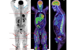
“As the vaccination program continues, we encountered increased F-18 FDG-activity not only in axillary lymph nodes ipsilateral to the injection site but also in other organs,” the authors noted.
To further elucidate cancer patient reactions to COVID-19 vaccines, the authors aimed to show any systemic metabolic changes after vaccination with three different vaccines in relation to time.
The group collected data on 220 eligible vaccinated cancer patients (127 with the Pfizer-BioNTech vaccine, 61 with the Moderna, and 32 with AstraZeneca vaccines) who underwent F-18 FDG-PET/CT scans. Of these, 71 patients also underwent a pre-vaccination scan. Most of the patients (n = 175 did not receive any therapy related to their diagnosis at the time of the PET/CT scan.
The researchers evaluated the exams from day 1 to day 135 after different vaccinations and compared the standardized uptake value (SUVmax) ratio of tracer activity of axillary lymph node to reference organs in all patients, with differences in tracer activity dynamics explored based on the three different vaccines.
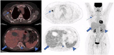
 A 73-year-old woman with a suspicious lung nodule was referred to our division for F-18 FDG PET/CT exam. She was vaccinated four days before PET/CT. The lung nodule did not show any pathological FDG uptake; however, high tracer activity was detected on her right arm, where she was injected, and multiple hypermetabolic lymph nodes in the right axilla (arrows). Moreover, PET/CT showed high tracer activity in the liver (black arrows), spleen (triangular arrows), and bone marrow (small triangular arrows) as the result of systemic immune response after vaccination. The tracer defect in the liver was due to the known cyst. (A) fusion scan, (B) PET, (C) maximum intensity projection. Image courtesy of Scientific Reports.
SUVmax of ipsilateral lymph node activity was slightly higher (mean, 3.00) in patients who received the Moderna vaccine than the BioNTech/ Pfizer-BioNTech (mean, 2.69) and AstraZeneca (mean, 2.61), according to the findings.
This FDG activity in axillary lymph nodes showed a steady decrease in all patients after Pfizer-BioNTech vaccinations. Ten days after vaccination, the FDG uptake was at its highest activity, and 70 days after vaccination, the FDG activity was not different from the background activity of the tracer in the axillary region, the researchers reported.
“This result also applies to other two vaccines,” the group wrote.
However, the authors highlighted several differences in terms of the timing of this activity. The highest peak of activity occurred in the fourth week for Moderna vaccinations and at the 10th day for AstraZeneca vaccinations.
Finally, the researchers noted that there was no significant changes of FDG tracer activity in other reference regions (the mediastinum, spleen, and bone marrow) between the three vaccinations.
Ultimately, numerous studies suggest increased activity in local sites and ipsilateral axillary lymph nodes in F-18 FDG PET/CT scans after COVID-19 vaccination, yet correctly interpreting these changes remains a challenge, the authors noted.
“Our study underscores the significance of changes in 2-[F-18] FDG PET/CT scans in lymph nodes and reference organs after vaccination and highlights the importance of this information in the interpretation of PET/CT in vaccinated patients,” the group concluded.
The full study is available here.


