A whole-body F-18 FDG-PET/CT scan was key in identifying medium-vessel vasculitis in a patient following a Moderna COVID-19 vaccination, a team in Australia has reported.
The diagnosis would have been difficult otherwise, given negative serology, and spared more invasive investigations such as muscle biopsy, noted nuclear medicine physician Martin Cherk, MD, of Alfred Hospital in Melbourne, and colleagues.
“F-18 FDG-PET/CT should be considered early as a potentially useful noninvasive investigation in patients suspected to have this condition following mRNA based COVID-19 vaccinations,” the group wrote. The case was published April 5 in EJNMMI Reports.
Although uncommon, a variety of autoimmune phenomena have been reported following COVID-19 infection or vaccination, including vasculitis, which is inflammation that can damage blood vessels and potentially lead to organ damage. These phenomena can be difficult to differentiate from other infective or inflammatory conditions, the authors explained.
While there have been several case reports of COVID-19 “BNT162b2” (Pfizer-BioNTech) and “mRNA-1273” (Moderna) vaccination-associated small- and medium-vessel vasculitis, none have used F-18 FDG PET/CT scans for the diagnosis, they noted.
In this case, a 57-year-old man presented with a two-week history of progressively worsening muscle pain, particularly around the thighs, and drenching night sweats, beginning three days following vaccination. He had two prior Astra-Zeneca ChAdOx1-S and two prior Pfizer-BioNTech COVID-19 vaccinations.
The patient had no cranial symptoms suggestive of giant cell arteritis, nor cardiorespiratory, otorhinolaryngeal, gastrointestinal, neurological or cutaneous symptoms suggestive of vasculitis. Blood tests demonstrated elevated inflammatory markers, while a neck and chest CT was normal.
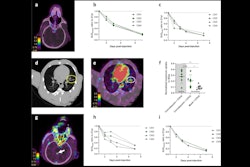
The subsequent F-18 FDG-PET/CT scan (Discovery 710, GE HealthCare) demonstrated extensive medium-vessel vasculitis throughout both thighs and to a lesser extent calves and proximal upper limbs.
 (A) Whole-body F-18 FDG-PET maximal intensity projection image demonstrating widespread medium vessel vasculitis predominantly involving both lower limbs (thighs and proximal calves) and much lesser proximal upper limbs/axillae (red arrows). Moderate FDG uptake in bilateral cervical nodes also demonstrated (blue arrows). Horizontal red lines depict different trans-axial slice levels. (B) Whole body F-18 FDG-PET/CT coronal fused images demonstrating medium vessel vasculitis involving bilateral femoral and popliteal vessels and numerous smaller branches throughout the thighs and proximal calves (red arrows). Horizontal red lines depict different trans-axial slice levels. (C) Whole-body F-18 FDG-PET/CT sagittal fused images demonstrating no abnormal FDG uptake in the ascending or descending thoracic aorta (blue arrow). Image and caption available for republishing under Creative Commons license (CC BY 4.0 DEED, Attribution 4.0 International) and courtesy of EJNMMI Reports.
The patient was started on daily prednisolone with rapid clinical and biochemical improvement and was discharged three days later. His myalgias, sweats, serum inflammatory markers, liver transaminases and hypoalbuminemia normalized within 21 days, according to the report.
“Given the striking F-18 FDG PET/CT scan findings typical for a widespread medium-vessel vasculitis and rapid response to treatment, the patient was not subject to biopsy and its inherent risks,” the group noted.
Ultimately, the report of a rare vaccination side effect does not diminish the established health benefits at a population level of COVID-19 vaccinations, including reducing mortality, the authors wrote.
The case demonstrates the excellent utility of F-18 FDG-PET/CT for the detection of medium-vessel vasculitis and exclusion of alternative diagnoses in this setting, Cherk and colleagues concluded.
The full case can be found here.


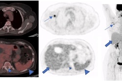


![A 53-year-old patient (patient number four) with a recurrent pituitary adenoma with extension of a cystic component of disease to the medial temporal lobe apparent on MRI (contoured in blue), and extension of disease to the left sphenoid bone and orbital apex apparent on [68Ga]Ga-DOTA-TATE (contoured in yellow).](https://img.auntminnie.com/mindful/smg/workspaces/default/uploads/2026/04/pituitary-tumor.QGsEnyB4bU.jpg?auto=format%2Ccompress&dpr=2&fit=crop&h=167&q=70&w=250)


