Clinical brain imaging with a newly developed portable PET scanner appears feasible, with a comparison to conventional PET imaging for the first time revealing similar results in human subjects, according to a group in New York City.
In a study in 20 healthy volunteers, the researchers found that a portable PET scanner measured brain metabolism based on F-18 FDG radiotracer uptake similarly to that of a standard PET scanner (Biograph mCT, Siemens Healthineers).
“The results obtained with a portable PET scanner in this comparison in humans require follow-up but lend confidence to the feasibility of more flexible and portable brain imaging with PET,” noted first author Elizabeth Bartlett, PhD, of the New York State Psychiatric Institute. The study was published on December 14 in the Journal of Nuclear Medicine.
Current PET scanners require patients to lie still during scanning and can only be used in centers large enough to support such a device, the authors noted. Conversely, recently developed portable PET scanners include seated or standing configurations and have been proposed for use in combat zones, sports fields, and intensive care units, the researchers added.
In 2018, the group received a grant from the U.S. National Institutes of Health to develop noninvasive approaches for estimating brain glucose metabolism using the new scanners. In this study, they aimed to establish the feasibility of the approach using an experimental scanner called CerePET (Brain Biosciences, Rockville, MD).
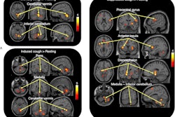
 A visual abstract describing a comparison between a portable PET scanner and standard PET scanner. Image courtesy of the Journal of Nuclear Medicine.
Each of the 20 healthy volunteers underwent dynamic F-18 FDG imaging with both scanners one to 154 days apart. Standard radiotracer uptake values (SUV) in brain tissue and rates of glucose metabolism (CMRglu) were quantified and compared between the scanners at regional and voxel levels.
According to the findings, outcome measures were well correlated, the group wrote. Specifically, correlation coefficients between the imaging sets across participants were 0.83 ± 0.07 for SUV and 0.85 ± 0.08 for CMRglu, the researchers reported.
“Our results indicate robust correlation and agreement between semi- and fully quantitative brain glucose metabolism measurements from portable CerePET and standard Biograph mCT scanners,” the group wrote.
Ultimately, at this stage, the major finding is that there appears to be no significant differences in fully quantified dynamic outcomes in cortical and subcortical brain areas between the two scanners indicating that the CerePET scanner is ready for imaging human subjects, they wrote.
“Future work will focus on more detailed scanner performance characterization, followed by application of CerePET to novel PET scanning scenarios,” the group concluded.
A link to the full study can be found here.