F-18 FAPI-PET/CT is superior to F-18 FDG-PET/CT for diagnosing and staging patients with pancreatic cancer, according to a study published January 4 in the Journal of Nuclear Medicine.
In a clinical trial, F-18 FAPI-04 radiotracer performed better than F-18 FDG in identifying primary tumors, lymph node metastasis, and distant metastases in patients with pancreatic ductal adenocarcinoma (PDAC). The findings suggest that F-18 FAPI-04 PET/CT offers a new method for identifying malignancies in these patients, according to first author Xiang Li, PhD, of Zhejiang University in Hangzhou, China, and colleagues.
“In the future, F-18 FAPI-04 PET/CT may play a greater role in the actual clinical management of PDAC,” the group wrote.
F-18 FDG is the most widely used cancer PET imaging radiotracer, yet its effectiveness for the detection and staging of suspected PDAC remains debatable, with studies suggesting its sensitivity ranges from 73% to 94%, according to the authors.
Conversely, FAPI radiotracers have been developed that target specific receptors in tumors. These tracers have shown promise in PDAC patients when labeled with gallium-68 (Ga-68) radioisotopes. Still, these Ga-68 tracers are short-lived, and preliminary research suggests FAPI tracers labeled with F-18 radioisotopes last longer, and thus may be delivered over longer distances and at a relatively lower cost, the researchers explained.
Moreover, the advantages of F-18 FAPI-04 over F-18 FDG have not yet been systematically evaluated in patients with PDAC, which is among the most lethal malignancies, they added.
To that end, the group enrolled 62 patients with confirmed PDAC at their hospital between August 2021 and February 2023. Patients underwent both F-18 FAPI-04 PET/CT and F-18 FDG- PET/CT at enrollment. After systemic treatment, response was evaluated bimonthly according to standard criteria, and final clinical staging was determined based on clinical, pathologic, and all imaging data.
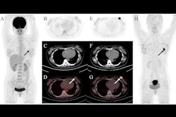
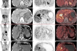
 Typical PET (top), PET/CT (middle), and CT and MR (bottom) images of primary tumor obtained using both radiotracers in representative patients (A and B). Tumor is marked by arrows. DWI = diffusion-weighted imaging. Image courtesy of the Journal of Nuclear Medicine.
F-18 FAPI-04 PET/CT identified all patients with PDAC, while F-18 FDG-PET/CT missed one patient, according to the findings. F-18 FAPI-04 tracer uptake was higher than F-18 FDG in primary tumors (10.63 vs. 2.87, p < 0.0001), lymph node metastasis (2.9 vs. 1.43, p < 0.0001), and distant metastases (liver, 6.11 vs. 3.1, p = 0.002; peritoneal, 4.7 vs. 2.08, p = 0.015).
In addition, 14 patients were upgraded and one patient was downgraded due to F-18 FAPI-04 PET/CT scans compared with F-18 FDG-PET/CT, the authors noted.
“Our results demonstrate that F-18 FAPI-04 PET/CT is significantly superior to F-18 FDG- PET/CT in detecting both primary and metastatic lesions,” the researchers wrote.
Ultimately, diagnosis and proper staging based on imaging assessments are essential for choosing treatment plans for tumor patients, and to that end, F-18 FAPI-04 PET/CT shows promise in PDAC patients, the authors wrote.
Future studies will analyze the correlation of F-18 FAPI-04 tracer uptake with survival outcomes after patients undergo treatment, which was beyond the scope of this trial, they noted.
“Additional studies are required to validate the prognostic value of F-18 FAPI-04,” the group concluded.
The full article can be found here.


![A 53-year-old patient (patient number four) with a recurrent pituitary adenoma with extension of a cystic component of disease to the medial temporal lobe apparent on MRI (contoured in blue), and extension of disease to the left sphenoid bone and orbital apex apparent on [68Ga]Ga-DOTA-TATE (contoured in yellow).](https://img.auntminnie.com/mindful/smg/workspaces/default/uploads/2026/04/pituitary-tumor.QGsEnyB4bU.jpg?auto=format%2Ccompress&dpr=2&fit=crop&h=167&q=70&w=250)


