PET imaging can help identify whether cardiac sarcoidosis patients are at higher risk of major adverse cardiac events (MACE), according to a study published August 7 in JACC: Cardiovascular Imaging.
In a meta-analysis of published cardiac PET studies, researchers found that abnormal FDG uptake and perfusion defect findings were particularly predictive.
“This study supports the routine use of FDG-PET in [cardiac sarcoidosis] for risk stratification given that patients who have specific features such as [right ventricular] uptake may be at higher risk of future MACE,” wrote lead author Tahir Kafil, MD, of the Cleveland Clinic in Cleveland, OH, and colleagues.
Cardiac sarcoidosis is characterized by the formation of granulomas in the tissue of the heart, and it can cause arrhythmias and heart failure. While there is no single diagnostic test for the disease (invasive biopsies are the most specific), PET imaging with FDG radiotracer is frequently used, as it identifies areas of cardiac inflammation. In addition, serial cardiac PET imaging is used to monitor how these patients respond to immunosuppression treatment.
While cardiac PET has been well studied for its diagnostic value, however, there is increasing evidence that it also provides important prognostic information, the authors explained.
To evaluate the evidence, the researchers searched current medical literature and pooled data from 55 cardiac PET studies involving 5,250 patients with cardiac sarcoidosis. The studies included reported outcomes on any cardiovascular adverse event, such as death, arrhythmia, ventricular tachycardia, ventricular fibrillation, heart block, cardiomyopathy, and heart failure.
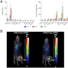
 A graphical abstract of the study. Image courtesy of JACC: Cardiovascular Imaging.
According to the findings, factors with the highest odds ratios (OR) associated with MACE included focal abnormal RV uptake (OR, 6.27), and a lack of response to immunosuppression on serial PET images (OR, 8.43).
Other risk factors included abnormal FDG uptake and perfusion defect (OR, 2.86), abnormal perfusion or FDG uptake (OR, 2.69), and abnormal FDG uptake (OR, 2.61), the researchers reported.
“Multiple cardiac PET parameters provide risk stratification value in cardiac sarcoidosis,” they noted.
Ultimately, the review findings contribute to the pool of published reports evaluating the prognostic capabilities of PET in cardiac sarcoidosis, the authors wrote. The analysis included 38 more PET studies and two more years of evidence than previous analyses.
Limits, however, included that most of the studies had a short duration of follow-up and small sample sizes, and longer-term evaluations are needed to assess whether FDG-PET remains prognostic, the team added.
“Although further studies are needed, overall, PET imaging provides multiple useful parameters for risk stratification,” it concluded.
The full study is available here.


![A 53-year-old patient (patient number four) with a recurrent pituitary adenoma with extension of a cystic component of disease to the medial temporal lobe apparent on MRI (contoured in blue), and extension of disease to the left sphenoid bone and orbital apex apparent on [68Ga]Ga-DOTA-TATE (contoured in yellow).](https://img.auntminnie.com/mindful/smg/workspaces/default/uploads/2026/04/pituitary-tumor.QGsEnyB4bU.jpg?auto=format%2Ccompress&dpr=2&fit=crop&h=167&q=70&w=250)


