


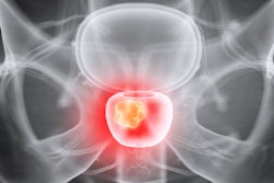
Insurance coverage for prostate MRI is inconsistent across the U.S., which may contribute to unequal access to the technology for patients, according to a study published September 7 in the American Journal of Roentgenology.
The difference appears to vary by facility, with different sites charging different amounts, wrote a team led by Dr. Aaron Brant of New York-Presbyterian Hospital in New York City.
"Patients may be responsible for paying for the examination but poorly equipped to compare highly variable charges among facilities," the team explained. "These concerns compound population disparities in insurance rates."
Although the use of MRI for prostate imaging is supported by professional guidelines, insurance coverage is variable, which translates to differences in out-of-pocket costs for patients, according to the authors.
"Uninsured or underinsured patients may be responsible for a significant portion of charges for prostate MRI, although this [fact isn't clear]," they wrote.
Brant and colleagues used information from the Premier Healthcare Database to investigate charge variations for prostate MRI exams conducted in U.S. hospitals between January 2010 and March 2020. The research included 552 facilities at which were performed 37,073 exams, and charges were adjusted to 2019 U.S. dollars. The team also tracked facility characteristics such as location (urban or rural and geographic region) and size.
Median charge per prostate MRI exam was $4,419, with a 26-fold range of $593 to $15,150. The team also found the following:
- Among patients paying for the exam themselves, median charge was $4,350, with a 25-fold variation of $550 to $13,815.
- Inflation-adjusted cost per prostate MRI exam increased from $4,192 in 2010 to $4,586 in 2020.
What factors contributed to the cost ranges? Variability between healthcare facilities was associated with 63.9% of charge inconsistencies; the next biggest factor was IV contrast, which was associated with 10.3% of the fee variations. Facility size, volume, geographic region, and patient characteristics had negligible impact, the team found.
Why variation among facilities was the primary factor for prostate MRI exam inconsistencies isn't clear, Brant and colleagues wrote.
"Potentially, absence of incentives to provide competitive prices enables hospitals to set arbitrary prices," they noted.
Cost variations may be compounded by higher patient payments due to out-of-network exams, "an increasingly common problem given the growth of narrow-network insurances," the team explained. And it also doesn't help that many hospitals don't comply with a mandate from the U.S. Centers for Medicare and Medicaid Services (CMS) to make charges for imaging exams public.
Potential fixes to prostate MRI cost variability do exist, the authors wrote.
"Expanded coverage, broadening of narrow networks, and stricter enforcement of price transparency regulations may ameliorate this problem," they concluded.


![Overview of the study design. (A) The fully automated deep learning framework was developed to estimate body composition (BC) (defined as subcutaneous adipose tissue [SAT] in liters; visceral adipose tissue [VAT] in liters; skeletal muscle [SM] in liters; SM fat fraction [SMFF] as a percentage; and intramuscular adipose tissue [IMAT] in deciliters) from MRI. The fully automated framework comprised one model (model 1) to quantify different BC measures (SAT, VAT, SM, SMFF, and IMAT) as three-dimensional (3D) measures from whole-body MRI scans. The second model (model 2) was trained to identify standardized anatomic landmarks along the craniocaudal body axis (z coordinate field), which allowed for subdividing the whole-body measures into different subregions typically examined on clinical routine MRI scans (chest, abdomen, and pelvis). (B) BC was quantified from whole-body MRI in over 66,000 individuals from two large population-based cohort studies, the UK Biobank (UKB) (36,317 individuals) and the German National Cohort (NAKO) (30,291 individuals). Bar graphs show age distribution by sex and cohort. BMI = body mass index. (C) After the performance assessment of the fully automated framework, the change in BC measures, distributions, and profiles across age decades were investigated. Age-, sex-, and height-adjusted body composition reference curves were calculated and made publicly available in a web-based z-score calculator (https://circ-ml.github.io).](https://img.auntminnie.com/mindful/smg/workspaces/default/uploads/2026/05/body-comp.XgAjTfPj1W.jpg?auto=format%2Ccompress&fit=crop&h=100&q=70&w=100)


![Overview of the study design. (A) The fully automated deep learning framework was developed to estimate body composition (BC) (defined as subcutaneous adipose tissue [SAT] in liters; visceral adipose tissue [VAT] in liters; skeletal muscle [SM] in liters; SM fat fraction [SMFF] as a percentage; and intramuscular adipose tissue [IMAT] in deciliters) from MRI. The fully automated framework comprised one model (model 1) to quantify different BC measures (SAT, VAT, SM, SMFF, and IMAT) as three-dimensional (3D) measures from whole-body MRI scans. The second model (model 2) was trained to identify standardized anatomic landmarks along the craniocaudal body axis (z coordinate field), which allowed for subdividing the whole-body measures into different subregions typically examined on clinical routine MRI scans (chest, abdomen, and pelvis). (B) BC was quantified from whole-body MRI in over 66,000 individuals from two large population-based cohort studies, the UK Biobank (UKB) (36,317 individuals) and the German National Cohort (NAKO) (30,291 individuals). Bar graphs show age distribution by sex and cohort. BMI = body mass index. (C) After the performance assessment of the fully automated framework, the change in BC measures, distributions, and profiles across age decades were investigated. Age-, sex-, and height-adjusted body composition reference curves were calculated and made publicly available in a web-based z-score calculator (https://circ-ml.github.io).](https://img.auntminnie.com/mindful/smg/workspaces/default/uploads/2026/05/body-comp.XgAjTfPj1W.jpg?auto=format%2Ccompress&dpr=2&fit=crop&h=167&q=70&w=250)


