Most clinicians would agree that mammography is an extremely effective way to find breast cancer. But it is a 2D modality used to image a 3D structure, a fact that has long been a thorn in the side of breast imagers.
"Mammography is still the most important modality to use for early detection," said Bonnie Rush, consultant and president of Breast Imaging Specialists of San Diego. "Nothing will take its place for the near future. But since it's a planar modality, it leads to excessive patient recalls because normal tissue is superimposed upon itself. There are other imaging technologies on the horizon intended to get around this problem."
Rush gave a primer on breast imaging technologies in various stages of clinical development and adoption at the American Healthcare Radiology Administrators (AHRA) annual meeting held this month in Las Vegas.
Digital breast tomosynthesis
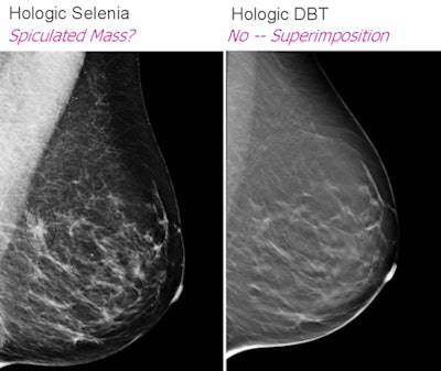
With its 3D lesion localization capability, digital breast tomosynthesis (DBT) can get around mammography's overlapping tissue problem, Rush said. Hologic (Bedford, MA), GE Healthcare (Chalfont St. Giles, U.K.), and Siemens Healthcare (Malvern, PA) all have work-in-progress units, and Hologic has submitted a premarket approval application to the U.S. Food and Drug Administration.
"Start planning for DBT in your budget," Rush told AHRA attendees. "It could be available next year."
With a DBT unit, both DBT and full-field digital mammography (FFDM) exams will be performed under a single compression, in craniocaudal and mediolateral oblique views. And although the radiation dose will not decrease, the exam will still require less radiation than is allowed in FFDM.
 |
| Slide courtesy of Bonnie Rush (images from Hologic). |
"FFDM plus DBT makes it easier to find changes in the breast than just FFDM alone," Rush said. "The combination has the potential to reduce screening recall rates by up to 40%. This will open up schedules and cut down on patient anxiety."
What are the unknowns for DBT? Technique optimization methods, the role of computer-aided detection, reimbursement, workflow timing, and real-life statistics, Rush said.
Conebeam breast CT
Conebeam breast CT (CBBCT) technology is still investigational, Rush said, and researchers are in the process of determining its value. As with DBT, it will potentially lessen the problems of superimposition of tissue and summation shadows that plague mammography.
CBBCT offers 360º views of breast anatomy, and the coned beam radiates only the breast, protecting surrounding tissue in the lungs, heart, and back. The patient lies prone on the exam table, so there is no compression. The investigational systems now installed show scan times of 10 seconds for low resolution and 48 seconds for high resolution, with 300 to 500 images per scan, Rush said.
A 2008 study found that mammography outperformed CBBCT for visualizing microcalcifications, but CBBCT was better than mammography for visualizing masses, Rush said. CBBCT could prove useful for imaging the postoperative breast and for image-guided surgery and interventional procedures. It also could prove equal or superior to breast MRI for staging tumors and screening high-risk women.
CT laser mammography
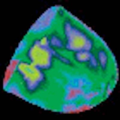
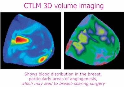
CT laser mammography (CTLM) examines blood flow to the breast, based on the idea that a tumor is more metabolically active than healthy tissue, and this metabolic activity can be detected by the laser technology. CTLM is not affected by tissue density and could become a way to determine response to therapy, according to Rush. Also, it's attractive to women because it doesn't require compression: Like CBBCT, the woman lies prone on a table with her breast hanging through an opening.
 |
| Slide courtesy of Bonnie Rush (images from Imaging Diagnostic Systems). |
But CTLM has some definite downsides, Rush said. Currently, it takes approximately 10 to 15 minutes to image each breast, even though the image reconstruction process occurs at the same time. In addition, data for the technology's sensitivity and specificity vary: In dense breasts, it beats mammography at 74.40% versus 34.4% for sensitivity, but comes in second for specificity at 72.2% versus mammography's 90.48%.
Breast MRI
Breast MRI has a high sensitivity rate -- but also a high false-positive rate -- and its specificity can be quite low, depending on the type of breast tissue. Studies have shown that breast MRI can slow the start of treatment by up to three weeks, and it drives an increase in prophylactic mastectomies.
"I do still call this an emerging modality," Rush said. "The concern is that breast MRI leads women to have more surgical intervention than needed and to delays in treatment."
Real-time virtual sonography
Real-time virtual sonography (RVS) can increase positive predictive value and decrease false positives in breast MRI, Rush said. It superimposes a sonography image with an MRI image of the same section in real-time and uses a position tracking system with a magnetic sensor. Its ability to detect incidental enhancing lesions is higher than with any other imaging technique.
"RVS can help radiologists diagnose potentially malignant lesions at an early stage to allow for more effective management of breast cancer," Rush said.
Molecular breast imaging and positron emission mammography
Molecular breast imaging and positron emission mammography (PEM) are not affected by dense breasts or implants. The imaging units are similar to mammography devices, and positioning requires an experienced technologist to produce the best results.
Molecular breast imaging uses a single gamma camera, and literature indicates that the presence of tumors smaller than 3 mm and/or located farther from the camera decreases the technology's sensitivity, Rush said. Another downside? The technology is vulnerable to vagaries in the supply of technetium-99m sestamibi.
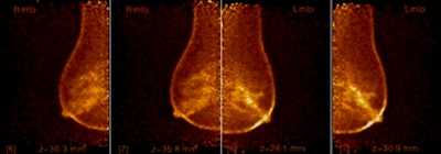
PEM, however, uses FDG, which is readily available. The system has two detector plates, allowing it to provide tomographic images, and scan time per breast is four to 10 minutes. PEM helps guide radiologic and surgical planning procedures, as well as monitor therapy. It also appears to be more specific in detecting ductal carcinoma in situ than MRI (73% versus 43%, respectively), according to Rush.
Molecular breast imaging's integration has been slow, because the communities of breast imaging and nuclear medicine aren't used to working together, Rush said.
As a complement to breast MRI, molecular breast imaging is cost-effective for radiodense breasts with indeterminate mammograms or ultrasounds, multiple regions of suspicion, or occult palpable masses. Its breast cancer indications include evaluating axillary regions for node status in breast cancer patients, treatment planning for multicentric or multifocal disease, and predicting chemotherapeutic response. The Food and Drug Administration has cleared molecular imaging as a problem-solving modality, while PEM is approved for patients with a current or past cancer diagnosis.
 |
| PEM depiction of ductal carcinoma in situ (DCIS). Forty-year-old premenopausal woman: PEM shows segmental uptake in left breast. Images courtesy of Naviscan. |
With all of the emerging breast imaging technologies to consider, clinicians are looking for reliable ways to find occult cancer, a particularly difficult task in dense breast tissue.
"It's what we can't see that kills," Rush said. "We have to identify the cancers before they can grow. And these advanced applications will continue to improve breast imaging."
By Kate Madden Yee
AuntMinnie.com staff writer
August 20, 2009
Related Reading
Study: MRI may cause more harm than good in breast cancer, August 13, 2009
Digital breast tomosynthesis cuts recall rate by 30%, August 4, 2009
CAD finds breast cancers missed at CR mammography, July 31, 2009
CARS report: Novel conebeam CT cuts breast dose, improves accuracy, June 29, 2009
CTLM with mammo boosts breast cancer diagnosis, December 4, 2008
Copyright © 2009 AuntMinnie.com


