Exposure to breast radiation therapy (RT) appears not to affect the prevalence of breast arterial calcifications (BAC) on screening mammography, according to research presented May 1 at the American Roentgen Ray Society (ARRS) meeting.
The finding is important because, since mammographic breast arterial calcifications can suggest the presence of cardiovascular disease (CVD), their being affected by RT would compromise this function, noted a team led by Jessica Rubino, MD, of Dartmouth Hitchcock Medical Center in Lebanon, NH.
"RT exposure is not associated with the presence of BAC and thus [BAC] can be utilized as an imaging biomarker for CVD risk in women," the group wrote.
Of women who present for screening mammograms, 10% to 15% have a personal history of breast cancer RT, Rubino and colleagues explained, writing that "understanding the association between breast arterial calcifications and breast radiation therapy is required for the appropriate utilization of BAC as a biomarker of cardiovascular disease risk."
To this end, Rubino's team investigated whether breast RT exposure affects the screening utility of BAC as an imaging biomarker of CVD via a study that consisted of an electronic health database query that identified 1,155 women between the ages of 40 and 75 who had a screening mammogram between January 2011 and December 2012 and no history of CAD. Data regarding any breast cancer radiation therapy history was removed; two breast imaging radiologists reviewed the mammograms for breast arterial calcification. The study authors assessed the association between breast RT exposure and BAC, adjusting for age, body mass index, smoking status, hypertension, type 2 diabetes, and use of statin and antihypertensive medication.
The group reported the following regarding the 1,155 women included in the analysis:
• 222 (19.2%) had mammographic evidence of BAC.
• 122 (10.6%) had a history of RT exposure.
• 39 (32%) women with a history of RT exposure had BAC on mammography.
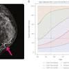
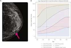
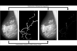
 Bilateral craniocaudal views show bilateral breast arterial calcification in a 71-year-old patient with history of left breast invasive lobular carcinoma treated with wide local excision and radiation therapy with 50.4 Gy and 10 Gy boost to tumor bed (scar market indicated dashed line). Mammogram obtained 4 years after completion of radiation therapy. Breast arterial calcification is more extensive in untreated right breast than in left breast that received radiation therapy. Image and caption courtesy of the ARRS.
The study indicates that BAC is not associated with RT exposure, Rubino and colleagues concluded -- which confirms that mammography can serve an additional purpose to identifying breast cancer.
"Universal reporting of breast arterial calcifications [identified on screening mammography] can provide sex-targeted screening that can improve cardiovascular disease risk stratification," the group noted. "This may lead to earlier preventive strategies and improved clinical outcomes."