Preoperative MRI does not yield better outcomes in women with human epidermal growth factor receptor 2 (HER2)-positive and hormone receptor-negative breast cancer, according to research published April 29 in Radiology.
In a retrospective study involving more than 1,000 women, researchers from the University of Ulsan College of Medicine in Seoul found that preoperative MRI was not associated with improvements in either recurrence-free survival or overall survival.
“Therefore, decisions regarding preoperative MRI should consider the overall clinical context of the patient rather than focusing solely on this cancer subtype,” wrote the team led by Hee Jeong Kim, MD, PhD.
As the long-term benefits and limitations of preoperative breast MRI for the HER2-positive and hormone receptor-negative invasive ductal carcinoma subtype haven’t been previously well-established in the literature, the researchers sought to assess the impact of the modality on long-term survival outcomes. They gathered data from 1,094 women with HER2-positive and hormone receptor-negative breast cancer receiving care at their institution between January 2007 and December 2016.
Of these women, 523 received preoperative MRI and 571 did not. Propensity score matching based on 19 clinical-pathologic covariates was used to account for various factors that could influence outcomes.
In the propensity score-matched set, preoperative MRI was not associated with recurrence:
What’s more, preoperative MRI was not associated with overall survival (HR: 0.63, p = 0.05).
After performing multivariable analysis, the researchers also found that preoperative MRI was not associated with improved recurrence-free survival (HR: 0.89, p = 0.44) or improved overall survival (HR: 0.73, p = 0.14).
“Further prospective multicenter studies with longer follow-up are essential to identify which patient groups are most likely to benefit from preoperative breast MRI, thereby ensuring its clinical relevance and cost-effectiveness,” the authors concluded.
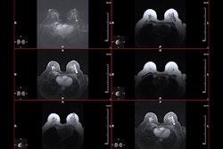
 Images show a palpable mass in the left breast in a 55-year-old woman. (A) Mammography shows a 26-mm ill-defined mass with fine linear calcifications in the central portion of the left breast (arrows). (B) ultrasound shows a 23-mm hypoechoic mass with calcifications in the corresponding area accompanying palpability (between the arrows). (C) An axial, fat-saturated, contrast-enhanced T1-weighted MRI scan shows an 18-mm irregular heterogeneous enhancing mass in the left breast (arrows). After breast-conserving surgery, the final pathologic report confirmed a grade II estrogen receptor–negative, progesterone receptor–negative, human epidermal growth factor receptor 2–positive invasive ductal carcinoma. There was no lymphovascular invasion, and the resection margins were clear. The patient declined both adjuvant chemotherapy and radiation therapy due to psychologic distress. (D) 11 months later, surveillance ultrasound helped to detect a new 25-mm irregular hypoechoic mass at the 12-o’clock position in the left breast (arrows). A subsequent ultrasound-guided biopsy confirmed it as a recurrence of grade II invasive ductal carcinoma, maintaining the same receptor statuses. Images courtesy of the RSNA.
In an accompanying editorial, Massimo Imbriaco, MD, and Andrea Ponsiglione, MD, of the University of Naples in Italy noted that the research clearly demonstrated that preoperative breast MRI has a minimal impact on long-term survival outcomes for women with HER2-positive and hormone receptor-negative breast cancer is minimal.
“Although there was a trend toward better [recurrence-free survival] and [overall survival] in the MRI group, these differences were not statistically significant in the matched cohorts,” they wrote. “Thus, the use of breast MRI in this particular group of patients should be reconsidered and avoided -- at least in patients with early-stage breast cancer to ensure that its use is both clinically relevant and cost-effective.”
The full study and accompanying editorial can be found here and here.

![Images depict axial MRI scans in a 50-year-old premenopausal female patient with biopsy-proven 12-mm invasive lobular cancer in the lower outer quadrant of the left breast. Upper images are subtracted dynamic contrast-enhanced (DCE) MRI scans from the first and second postcontrast acquisitions. Lower images are ultrafast MRI scans obtained within 24 hours after the DCE study (four consecutive subtracted dynamic frames from time points [T] 6–9). There is marked background parenchymal enhancement (BPE) on the DCE MRI scans. Thus, the known cancer (arrow) is barely visible; it exhibits only slightly stronger enhancement than the normal fibroglandular tissue, which results in low conspicuity. At ultrafast MRI there is less BPE, and still the cancer is barely visible because it starts to enhance simultaneously with the BPE. The conspicuity of the cancer is even reduced compared with the DCE series. Both of the readers missed this cancer on both the DCE and ultrafast MRI scans.](https://img.auntminnie.com/mindful/smg/workspaces/default/uploads/2025/05/2025-05-06-rsna-ultrafast-dce-mri-breast.Gzvq76udFr.jpg?auto=format%2Ccompress&fit=crop&h=167&q=70&w=250)