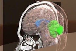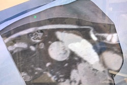
Augmented reality (AR) technology and 3D CT holograms can significantly improve recall of head and neck anatomy in first-year medical students, yielding better performance than viewing the holograms on traditional laptop displays, according to research published online August 20 in Academic Radiology.
A team of researchers from the University of Pennsylvania led by Dr. Joanna Weeks provided a review of head and neck anatomy in a teaching session to two groups of 15 first-year medical students -- one group utilized an AR headset and advanced visualization software, while the other viewed the hologram using the same software on their 2D laptop screens.
Although both groups answered significantly more questions correctly on a five-question anatomy test after the session than they had beforehand, the group that employed AR headsets achieved the best results.
"Immersive 3D visualization has the potential to improve short-term anatomic recall in the head and neck compared to traditional 2D screen-based review, as well as engage millennial learners to learn better in anatomy laboratory," the authors wrote.
Hypothesizing that the benefits of stereoscopic depth cues provided via AR software would produce better results than 2D displays, the researchers first created a 3D CT hologram from a noncontrast-enhanced CT exam performed on a donated cadaver. The 15 students in the AR group then viewed the hologram using a Microsoft HoloLens with the SurgicalAR (Medivis) AR 3D image visualization software. Meanwhile, the 15 students in the laptop screen group utilized SurgicalAR on a Dell laptop display.
| Test performance before and after head and neck anatomy teaching session using 3D CT hologram | ||
| Group using laptop screens | Group using AR headset | |
| Average presession percentage of correct answers | 57% | 59% |
| Average postsession percentage of correct answers | 80% | 95% |
Although the performance difference after the teaching session was statistically significant in both groups (p < 0.05), the results were significantly better in the AR group (p = 0.022).
"Our findings may reflect additional benefit gained from the stereoscopic depth cues present in augmented reality-based visualization," the authors wrote.
In qualitative feedback, both groups of participants reported that their perceived understanding of anatomic relationships had significantly improved after the session, according to the authors. And although the researchers didn't find a statistically significant difference between the two groups for this improvement, the AR group generally "provided lengthier and more varied responses and used more emphatic language than the screen group."
AR offers advantages over 3D physician models, which, though useful for training, are costly to obtain and store and can't readily be used to teach radiologic-anatomic correlation, according to the researchers.
"Augmented reality platforms may allow educators to create an immersive experience similar to viewing a physical model but allowing for radiologic overlays and direct radiologic-anatomic correlation," they wrote.
This would require medical schools or radiologic education departments to invest in an AR-capable headset and the software to display the CT data. However, these costs could partially be offset by the use of "near-peer" instructors such as senior medical students or residents.
"Near-peer instructors are favored by many millennial learners and could additionally facilitate an increased instructor-to-student ratio," they wrote.

