Dear Advanced Visualization Insider,
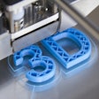
3D printing can be a valuable tool for preoperative planning. In a recent study, a team led by researchers from Florida Atlantic University found that a 3D-printed replica of a spine based on a CT exam could potentially facilitate improved results for cervical disk implants in patients with degenerative disk disease.
After modifying a 3D-printed spine replica with an artificial disk implant and a flexible magnetic sensor array, the researchers found that artificial intelligence (AI) algorithms could then accurately detect applied loads, as well as postures of the spine. As a result, surgeons could potentially utilize these personalized spine "twins" to preview and compare potential interventions prior to surgery. You can learn all about this research by reading this edition's Insider Exclusive.
In other 3D printing news, the combination of percutaneous nephrolithotomy and 3D-printed models produced from patient CT scans led to significantly better treatment of complex kidney stones, according to a recent study. AI could also increase productivity in 3D labs.
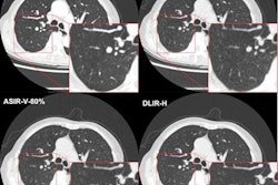
Also, an image reconstruction algorithm based on deep learning was found to enable detection of more lung nodules on ultralow-dose CT exams.
The utility of radiomics was also featured in a number of recent studies. For example, the pairing of CT radiomics and machine learning was able to predict survival of melanoma patients after immunotherapy better than relying on the Response Evaluation Criteria in Solid Tumors (RECIST) 1.1 guideline. A few hurdles still remain, however, before the technique could be applied in clinical practice.
In addition, a nomogram that utilizes CT radiomics data can preoperatively predict lymph node metastasis in patients with pancreatic ductal adenocarcinoma. Machine-learning models based on mammography radiomics also helped predict which patients with ductal carcinoma in situ (DCIS) should be upstaged. These algorithms outperformed models trained using only clinical criteria.
When it comes to performing brain volumetry on an ultrafast 3D brain MRI sequence, conventional image acquisition still yields the best results, researchers recently reported. And augmented reality visualization of SPECT/CT data can help to guide surgeons in identifying sentinel lymph nodes in patients with metastatic melanoma.
Is there a story you'd like to see covered in the Advanced Visualization Community? Please feel free to drop me a line.


