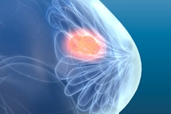
A patient presents with a high RENAL score tumor (≥8): a deeply endophytic renal lesion, invisible on the kidney surface, and located close to critical vasculature. The surgical goal is clear: remove the tumor while preserving as much healthy kidney tissue as possible. But achieving that goal depends entirely on understanding anatomy that cannot be fully appreciated on 3D volume rendering alone.
 Beatriz Dominguez Gonzalez, global market manager, Materialise
A 2022 systematic review published in Kidney Cancer highlights the clinical impact: "The application of 3D models for preoperative planning has been reported to increase the selective clamping rate and reducing the opening of collecting system, blood loss, and loss of renal function." The challenge is consistent: Complex spatial relationships are difficult to visualize accurately from volume rendering alone, a challenge that 3D surface modeling is designed to address.
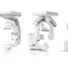
In complex partial nephrectomy cases like these, radiology is no longer limited to describing tumor size and location. Increasingly, radiologists are converting CT and MR datasets into patient-specific 3D surface models that clearly delineate healthy parenchyma, tumor margins, vascular supply, and the pyelocaliceal system.
These models allow surgical teams to visualize spatial relationships, evaluate selective arterial clamping strategies, and anticipate ischemia risks before entering the operating room. For high-complexity tumors, that level of anatomical clarity can influence whether radical resection or an organ-sparing approach is safely attempted.
This is where the evolving role of radiology becomes visible. As minimally invasive surgery and precision medicine advance, imaging is expected to do more than confirm diagnosis. It is expected to support surgical decision-making in a way that directly impacts procedural confidence and patient outcomes.
Structured 3D workflows embedded within radiology departments are increasingly becoming part of routine care. Not as an experimental adjunct, but as an established, evidence-supported standard that is raising the bar for surgical predictability.
From volumetric data to actionable 3D insight
The technical foundation already exists. CT and MRI are inherently 3D modalities, and radiologists interpret volumetric datasets every day. Most routine workflows rely on volume rendering within PACS, optimized for diagnostic efficiency rather than surgical planning, which requires interactive models that surgical teams can manipulate and explore.
3D surface modeling provides the interactivity needed for surgical planning. Radiology teams segment specific structures and convert them into discrete 3D objects, digital models of bones, vessels, organs, and pathology. These models can be rotated, isolated, highlighted, or hidden to reveal underlying structures, and explored spatially, enabling surgeons to test approaches and plan procedures in ways that volume rendering cannot support.
The clinical applications are concrete. In orthopedics, 3D surface models support trauma and fracture planning, allowing surgeons to assess fracture patterns and virtually restore bone alignment before the operation, surpassing the capabilities of 2D x-ray imaging. In cardiac interventions, they enable precise structural visualization for device sizing and placement planning. In cranio-maxillofacial surgery, they enable precise planning of orthognathic procedures, aligning skeletal structures and allowing soft-tissue simulation to improve bite function and facial symmetry. In oncology, they clarify tumor-vessel relationships in organ-sparing procedures, as in the nephrectomy case.
The underlying principle is consistent: transforming imaging from descriptive to spatially interactive. This approach has gained traction at leading institutions. More than 500 hospitals worldwide have integrated in-house 3D planning with 3D surface modeling into their surgical preparation workflows, with published data from institutions like Mayo Clinic documenting benefits in operating time and procedural predictability.
Embedding 3D as a standard radiology workflow
A common misconception is that 3D planning with surface modeling requires a physical 3D print lab or large capital investment. In reality, much of the clinical value derives from digital 3D surface models rather than printed objects. Interactive 3D visualization, shared during planning sessions or via augmented reality platforms, provides dynamic anatomical insights without requiring dedicated print labs.
When 3D surface modeling is embedded within the radiology department, it offers distinct operational advantages: direct control over turnaround times and quality standards, seamless integration with existing imaging workflows, access to subspecialist expertise within the institution, and the ability to tailor protocols to specific surgical service needs. For departments managing growing case volumes and complexity, keeping this capability in-house provides both clinical flexibility and cost predictability.
Most successful programs follow a pragmatic pathway: Identify surgical services with high-complexity cases, standardize segmentation protocols for defined indications, align turnaround times with surgical scheduling, and maintain quality control within radiology. Importantly, 3D surface modeling serves as the foundation for all downstream applications, whether that's digital planning, augmented reality visualization, or physical 3D printing of anatomical models and surgical guides. The digital models themselves deliver clinical value immediately, while printing remains an option when tactile feedback or sterilizable guides are required for specific cases.
In the United States, formal recognition for 3D surface modeling is now in place. In September 2025, the AMA CPT Editorial Panel approved six new Category III codes for digital 3D surface modeling workflows: surface modeling (1030T), surgical simulation (1032T), and computational modeling (1034T), with additional three codes for extended procedural time. Effective July 2026, these codes establish a pathway for reimbursement and a framework for radiology departments to adopt these workflows and capture both clinical and financial benefits without requiring additional investment in 3D printing infrastructure.
Radiology's expanding clinical footprint
In conversations with radiology departments that use 3D planning, one theme consistently emerges: These tools fundamentally change how radiologists and surgeons communicate. When both teams interact with the same patient-specific 3D model, the radiologist's understanding of imaging artifacts, anatomical variants, and structural relationships becomes directly accessible in a visual format that surgeons can manipulate.
Surgical approaches can be tested against patient anatomy before entering the operating room. Potential complications surface during planning rather than during the procedure. This shared visual language reduces the friction that can exist between diagnostic and procedural teams, creating stronger interdisciplinary partnerships.
3D surface modeling does not replace diagnostic interpretation. It extends it in two important ways. First, it reveals key anatomical details and relationships with a clarity and spatial precision that volume rendering alone cannot provide. Second, it transforms communication between radiology and surgical teams by creating a common visual language, converting complex data into interactive models that surgical teams can manipulate and explore together in real time, rather than mentally reconstructing.
From a technology provider's perspective, the pathway forward is increasingly clear. 3D surface modeling software is integrating better with PACS workflows. Reimbursement structures are emerging to support this work. Clinical evidence continues to accumulate from leading institutions.
Radiology has always shaped clinical decisions. Structured 3D surface modeling workflows raise the bar, making the connection between imaging intelligence and procedural outcomes more direct and visible.
Beatriz Dominguez Gonzalez is global market manager at Materialise.
The comments and observations expressed are those of the author and do not necessarily reflect the opinions of AuntMinnie.com.