A machine-learning analysis of functional MRI (fMRI) scans has shed light on key brain regions that differentiate superagers from cognitively average older adults, according to a poster study to be presented May 15 at the International Society for Magnetic Resonance in Medicine (ISMRM) annual meeting in Honolulu, HI.
Specifically, the study used a random forest machine-learning algorithm (RF-MLA) and identified the salience network, the default mode network, and the executive control network right -- three of six core networks involved in cognitive function -- as pivotal networks for superager classification.
“While the use of advanced analytical techniques and whole-brain approaches validates existing findings, we also observed new insights into sensory cortices that may be important for comprehending cognitive resilience in aging,” noted lead author Laiz Laura de Godoy, MD, PhD, of the University of Pennsylvania in Philadelphia, and colleagues.
Superagers are older adults with episodic memory performance similar or superior to middle-aged individuals. Previous research in predicting superagers using resting-state fMRI have largely concentrated on well-established networks linked to cognition and overlooked other key brain hubs, the researchers explained.
To address the knowledge gap, the group developed a data set based on a cohort of 31 older adults, 80 years or older, who were grouped as superagers (n=14) or age-matched healthy older adults (n=17). They used an RF-MLA first to phenotype superagers in six preselected cognitive networks: the default mode network (DMN), the executive control network right (ECN-R), the executive control network left (ECN-L), and the hippocampal, language, and salience networks (SN). They also compared findings with their previous work, in which they used an Elastic Net regression machine-learning model on the same networks and cohort.
Next, they expanded the superagers phenotyping and explored whole-brain connectivity across 11 distinct networks, including the six networks used in first analysis, plus the auditory, sensorimotor, visual lateral, visual medial, and visual occipital networks.
“Across these networks, we derived the most important nodes within the most relevant networks and hence identified brain regions that differentiate superagers from cognitively average age-matched healthy older adults,” the group wrote.
According to the results, in both the preselected and whole-brain network analyses, the key networks and nodes that differentiate superagers and age-matched healthy older adults were similar to their previous work. Specifically, the RF-MLA identified the SN, DMN, and ECN as pivotal networks for superager classification, with particular importance for the SN in the present study.
Moreover, the RF-MLA determined additional nodes in the auditory, visual-lateral, and visual-medial networks pertinent to superagers and highlighted the discriminative value of the visual-lateral network, the researchers noted.
“Our study utilized an RF-MLA to shed light on the significance of key networks and nodes in characterizing superagers,” the group wrote.
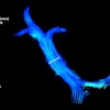
 A lollipop plots showing the most important nodes by RF-MLA selected by Mean decrease in Gini for a 7 tesla fMRI dataset, with longer sticks meaning greater importance. Each group of networks’ nodes has its own color matching the same network’s color in the brain map. Nodes' names preceded by a red asterisk are repeated among different networks, matching the red nodes in the brain map, which appear in multiple networks.Laiz Laura de Godoy, MD, PhD, and the ISMRM
Ultimately, the study validates existing findings and gives new insights into sensory cortices that may be important for superagers, according to the researchers.
“Although further research is needed in larger prospective studies, these findings might be helpful to guide future targeted interventions to optimize the efficiency of specific brain regions,” the group concluded.
Check out AuntMinnie.com’s full coverage of ISMRM 2025 here.