Dear Musculoskeletal Imaging Insider,
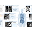
A new study from St. Jude Children's Research Hospital in Memphis, TN, found that routine bone scans may be safely eliminated and replaced with FDG-PET/CT in the evaluation of higher-risk children, adolescents, and young adults with Hodgkin's lymphoma.
The research, which was presented at the recent annual SNM meeting in New Orleans, found that all abnormal foci of uptake seen on bone scintigraphy representing metastatic disease also were evident through FDG-PET/CT scanning.
More details and clinical images from this Insider Exclusive are available by clicking here.
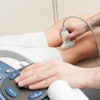
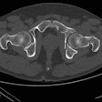
Also in this edition is the case of a 17-year-old soccer player who injured himself while slowing to kick the ball during a game -- he slipped and experienced immediate pain in his right thigh and hip.
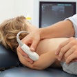
The case study discusses how ultrasound was used to image the rectus femoris muscle and explains why the muscle is particularly vulnerable to eccentric stress forces, such as the contractions associated with sprinting and jumping. The report's authors are from the Studio Ecograficao Dott. Stefano Ciatti in Prato, Italy.
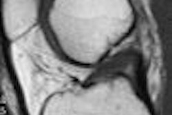
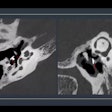
We're also featuring a piece on computer-aided detection (CAD) technology of knee MRI studies and its utilization in the automated detection of meniscal tears. Staff writer Erik Ridley writes how the performance of this CAD application was comparable to that of board-certified specialty-trained radiologists for automated detection of meniscal tears, and how CAD has gained acceptance for certain image interpretation tasks.
Keep in touch with the Musculoskeletal Imaging Digital Community in the coming weeks as we report on more late-breaking news and research from a variety of sources around the world.
