General Gallium 67 Tumor Imaging:
Chemistry and Pharmacology:
Gallium 67 is produced in a cyclotron by bombarding a zinc target
with protons. It has
a physical half-life of 78 hours and a biologic half-life of 2 to 3
weeks. Ga-67 decays by
electron capture to ground state Zn-67 with the following gamma
emissions: 93 keV(40%),
184 keV(24%), 296 keV(22%), 388 keV(7%). The 388 keV emission is
generally not used for
imaging. Gallium has approximately a 12 hour half-life in blood. The
Ga+3 ion resembles
the ferric (iron) ion in atomic radius and charge and binds to
transferrin (in plasma) [1]. The gallium transferrin complex binds to
transferrin receptors and is incorporated intracellularly where it
binds to
lactoferrin and siderophores (in tissue) [1]. The agent also binds to
ferritin.
That which is not protein bound is either cleared by the kidneys or passes into the extravascular space. Saturation of transferrin with iron, as seen clinically in patients with iron overload from repeated transfusions, will result in altered biodistribution of gallium with less liver uptake, greater renal activity, and more rapid clearance. Renal excretion (10-30% of injected dose) occurs mostly during the first 24 hours. This is because gallium citrate is excreted differently than gallium which is bound to transferrin. Renal activity should NOT be seen on 72 hour images. Persistent activity implies renal disease (ATN, renal insufficiency), pyelonephritis, inflammatory nephritis, or severe hepatic failure. Gastrointestinal excretion is the predominant route after 24 hours, primarily through the mucosa of large intestine, but a small amount is cleared via the liver and biliary tract. A laxative may be required prior to later imaging to clear colonic activity.The injected radiotracer must be "carrier free", ie: free of stable gallium. Significant amounts of carrier gallium will change the biologic distribution of Gallium 67 in the body, resulting in increased concentration in the skeleton.
Distribution:
Gallium activity can be seen normally in the following locations:
- Renal cortex: First 24 hours
- Liver: Demonstrates greatest uptake of gallium
- Spleen
- Bone marrow & skeleton:
- Gallium is incorporated into the calcium hydroxyapatite crystal as a calcium analog. Marrow activity occurs because of its behavior as an iron analog.
- May see physeal activity in children
- Nasopharynx, salivary & lacrimal glands
- Bowel: Primarily colonic activity on delayed images
- Breasts: Uptake is especially noted under the stimulation of menses, pregnancy, or BCP's. Gallium also accumulates in breast milk.
- External genitalia
- Blood pool (20%)
- Thymus: May note thymic uptake in children
Gallium Imaging Technique:
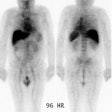
The typical dose of Gallium is 10 mCi for neoplasms (Image at 48-72
hours). When
performing a total body scan, a slow scan speed (10 cm/min.) is
recommended in order to
obtain adequate count densities. Spot images should be obtained for 10
minutes or 750,000
counts using three gallium peaks. When performing spots of the abdomen,
the liver should
be shielded or placed out of the field of view. Concurrent TcSc imaging
may be performed
to subtract out liver activity. When imaging axillary abnormalities,
the arms should be
raised above the head to fully expose the axilla.
REFERENCES:
(1) AJR 2011; Kashefi A, et al. Molecular imaging in pulmonary
diseases. 197: 295-307
