Bone Trauma:
Injuries of the Thoracic Spine:
Clinical:
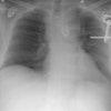
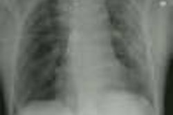
Fractures of the thoracic spine occur in 3% of blunt trauma cases [2]. Thoracic spine fractures are often associated with sternal fractures [2].The most common post-traumatic fractures seen in the thoracic spine are anterior wedge compression fractures and burst fractures. An anterior compression fracture is usually stable, while a burst fracture is unstable. The majority of fractures occur near the thoracolumbar junction (T9-T11) [2]. Nearly 62% of patients with thoracic spine fractures have complete neurologic deficits. The high incidence is likely secondary to the narrow diameter of the thoracic spinal canal and the potential for compromise to the vascular supply to the middle portion of the cord.X-ray:
The thoracic vertebral bodies are normally slightly wedge shaped- however, the posterior height should not exceed the anterior height by more than 2 mm. Between 70-90% of spine fractures can be identified on plain radiographs, but may be difficult to identify on portable, AP exams. In one study, only 51% of spinal fractures were identified at initial chest radiography [2]. Findings include widening of the paraspinal lines, mediastinal widening, and loss of vertebral body height [2]. If several posterior ribs seem to emanate from the same vertebral body ("crab sign"), this can be a clue to a thoracic spine fracture. CT is more sensitive to the detection of thoracic vertebral fractures than plain film. MRI has been very useful in demonstrating the presence of cord injury in patients without radiographic abnormality (SCIWORA- spinal cord injury without radiographic abnormality syndrome).Rib Injury:
(See also "Flail Chest")Clinical:
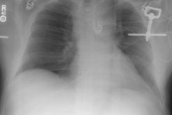
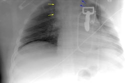
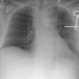
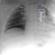
The most common skeletal injury in blunt chest trauma is rib fracture [3]. Rib fractures are found in about 10-50% of patients admitted for trauma [1,2,3], however, because not all rib fractures are seen on radiographs, the actual incidence is probably higher (approaching 60% of patients [2]). In fact, the CXR may underestimate the presence and extent of rib fractures by as much as 50% [1]. Although rib fractures can be quite painful, they are generally on little clinical significance and treatment is typically conservative. Displaced fractures of the lower ribs may perforate the lung, liver, or spleen [3]. Fractures of the 1st through 3rd ribs are considered to be associated with high energy trauma and they can be associated with brachial pleuxus and subclavian vessel injuries [3], as well as bronchial tear or aortic laceration..Other Fractures:
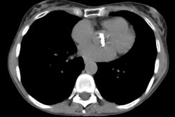
Sternal fracture: Motor vehicle accident is the most common cause of sternal fracture- the prevalence of sternal fracture among patients with blunt chest trauma ranges from 3-7% [4]. Most fractures occur in the upper or mid-body and the manubrium [3]. Blunt cardiac injury can be identified in 18-62% of patients with sternal fracture [1]. Clinicians should maintain a high index of suspicion for cardiac injury in patients with sternal fracture and consider ECG and cardiac rhythm monitoring for 12 hours [1].Sternoclavicular dislocation: Anterior dislocation is more common than posterior [3]. Anterior dislocation typically have a more benign course and are treated conservatively [3]. Posterior dislocation are more serious as they can result in injuries to the mediastinal vessels, trachea, or esophagus [3]. A posterior dislocation may be subtle or not visualized on CXR [3].
Scapular fracture: Scapular fractures are uncommon and indicate a high energy trauma [3]. Scapular fracture is associated with an increased risk for pulmonary contusion (54%), rib fractures (51%), and ipsilateral subclavian, axillary, or brachial artery injury (11%).
REFERENCES:
(1) Chest Surg Clin N Am 1997; Mayberry JC, Trunkey DD. The fractured rib in chest wall trauma. 7 (2) May: 239-260
(2) Radiographics 1998; Kuhlman JE, et al. Radiographic and CT findings of blunt chest trauma: Aortic injuries and looking beyond them. 18: 1085-1106
(3) Radiographics 2008; Kaewlai R, et al. Multidetector CT of blunt thoracic trauma. 28: 1555-1570
(4) Radiographics 2009; Restrepo CS, et al. Imaging appearances of the sternum and sternoclavicular joints. 29: 839-859
