Myocardial Contusion:
Clinical:
Myocardial contusion occurs in 8 to 71% of patients following severe blunt trauma. In autopsy series, cardiac contusion is found in 16% of patients who died following chest trauma. Cardiac contusion most commonly nvolves the anterior surface of the heart (injuries to the right ventircle are twice as common as LV injuries) [3]. Although the injury is usually not clinically significant and is difficult to diagnose, complications such as arrhythmia, acute valvular regurge, ventricular dysfunction, or rupture can occur. Additionally, patients with cardiac contusion are typically more severely injured. Rib fractures are found in 83% of patients with cardiac trauma, pneumothorax in 39%, hemothorax in 31%, lung contusion in 13%, clavicular fracture in 13%, and sternal fracture in 10% [3].Findings which suggest the diagnosis include chest pain, a pericardial friction rub, and an S3 gallop. On ECG there are typically non-specific ST-T changes, although a conduction abnormality or ST segment elevation in two contiguous leads may be seen. The presence of dysrhythmias in a chest trauma patient should indicate cardiac contusion until proven otherwise [1]. Elevated CK-MB levels are highly sensitive for cardiac injury (>90%), but can be elevated in trauma patients the absence of cardiac injury and lack specifity (<6.1%) [3]. The presence of a negative ECG and normal CK-MB levels have a very good negative predictive value for the exclusion of cardiac injury [1]. Other biomarkers such as troponin T, and troponin I are both sensitive and specific (>90%) for detection of cardiac ijury and their level of elevation is proportional to the extent of myocardial damage [3].
X-ray:
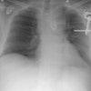
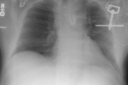
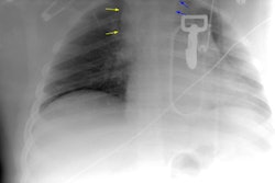
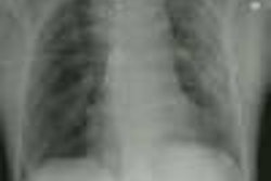
The plain film can demonstrate evidence of trauma to the thoracic cage and findings of heart failure.Thallium defects on scintigraphic imaging can be seen in areas of myocardial contusion, but perufison imaging is typically not helpful as most contusions involve the right ventricle which is not well imaged by scintgraphy [3]. The reported sensitivity is 55%, specificity 32%, and accuracy 38% [3].
On cardiac echo, segmental wall motion abnormalities can be identified in the involved areas [3]. The involved segments also demonstrate increased echogenicity [3].
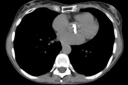
On CT, the presence of a hemopericardium is suggestive of myocardial rupture, aortic root innjury, or coronary artery laceration [3].
REFERENCES:
(1) Chest 1995; Feghali NT, et al. Blunt myocardial injury. 108:
1673-77
(No abstract available)
(2) Radiographics 2008; Kaewlai R, et al. Multidetector CT of blunt
thoracic trauma. 28: 1555-1570
(3) Radiographics 2012; Restrepo CS, et al. Imaging patients with
cardiac trauma. 32: 633-649
