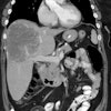
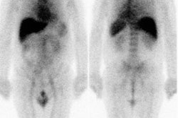
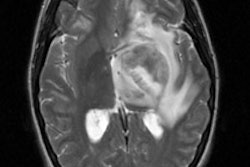
This is a case where CEA-Scan finds occult recurrence missed by both CT and dedicated FDG-PET. The patient has a rising serum CEA, and was otherwise free of symptoms of recurrence.
PATIENT HISTORY: A 44 year-old female with right colon cancer (mucinous carcinoma) resected in May 1998 followed by a hysterectomy in December of 1998. Chemotherapy started in August of 1998 continues at the time of this evaluation.
SERUM CEA LEVEL: 38 ng/ml
PRIOR DIAGNOSTIC STUDIES:
Negative
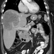
abdominal/pelvic CT (May 1999)
Negative
abdominal/pelvic PET (June 1999)


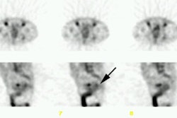
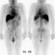
CEA-Scan FINDINGS: SPECT Imaging (June 1999) identifies focal activity in the right pelvis superior to the bladder (black arrow).
SURGICAL FINDINGS: Metastatic disease in the right pelvis.
CONCLUSION: CEA-Scan can change patient management by identifying the source of rising serum CEA levels missed by both CT and PET.
CEA-Scan was performed by Dr. Josef Machac on a Hitachi camera at Mount Sinai Medical Center, NY.
Additional information regarding mucinous adenocarcinomas: This false negative case may be explained by the impairment of glucose uptake by mucinous tumors, due to their hypocellularity. A study published last year by Dehdashti et al (Dis Colon Rectum 2000 Jun;43(6):759-67) reported that dedicated FDG-PET has a sensitivity of 92% for non-mucinous recurrent colorectal cancer, but is only 58% sensitive for mucinous tumors. Mucinous tumors comprise about 18% of the colorectal cancer population, or approximately 120,000 people who are being followed for recurrence.
