CNS Neoplasms:
Primary CNS neoplasms are uncommon (7 to 19 cases per 100,000 people) [9]. Brain metastases are up to 10 times more common than primary brain tumors and occur in 20-40% of patients with cancer [9]. The most common tumor types to metastasize to the brain are lung, breast, and melanoma [29].
Primary CNS gliomas are of 3 main types- astrocytomas,
oligodendrogliomas (more likely to contain calcification and have
a better overall prognosis), and mixed oligoastrocytomas [29]. The
tumors are further classified as low grade (WHO grades I and II)
or high grade (WHO grades III and IV) based upon the degree of
nuclear atypia, mitoses, microvascular proliferation, and necrosis
[29]. There are 3-subtypes of low grade gliomas- pilocystic
astrocytoma (grade I), astrocytoma (grade II), and
oligodendroglioma (grade II) [29]. Low-grade (grade II) gliomas
can have a very variable clinical course [53]. Some patients
demonstrate indolent disease, while other progress rapidly to
high-grade gliomas [53].
High grade gliomas include anaplastic tumors (grade III) and glioblastoma (grade IV) [29]. GLioblastoma is the most malignant and most common glioma- accounting for 45-50% of cases [29]. The mean age of onset for glioblastoma is 61 years, and 40 years for anaplastic astrocytoma [29]. The clinical course of glioblastoma is usually rapid and fatal with a median survival or 1 year [29]. The median survival for anaplastic tumors in 2-3 years [29]. Low-grade tumors are more commonly seen in younger patients [29].
Treatment for grade III and IV tumors is surgical resection to
the most feasible extent, followed by adjuvant radiotherapy to
improve survival [29]. Concurrent chemotherapy with temozolomide
has also been shown to improve survival in patients with
glioblastomas [29].
Bevacizumab (Avastin) is a humanized monoclonal antibody
inhibiting the biologic activity of vascular endothelial growth
factor and the agent is increasingly used for the treatment of
patients with recurrent high-grade gliomas- either as a single
agent or in combination with irinotecan (a topoisomerase 1
inhibitor) [51]. Differentiation of tumor response from
progression can be made difficult by apparent non-enhancing tumor
progression and pseudo-response (pseudo-regression) as a result of
drug-induced normalization of tumor vasculature [63,68].
Anti-angiogenic agents (like bevacizumab) can reduce contrast
enhancement as early as 1-2 days (due to normalization/restoration
of the blood-brain barrier, rather than actual reduction in the
lesion), resulting in apparent spuriously high response rates of
up to 60% [68]. Steroids can also result in reduction in tumor
size on MRI [68].
Low grade gliomas are more indolent, but can still be fatal lesions [29]. For pilocytic astrocytomas, surgical resection alone leads to a cure or long-term survival (20 year survival of 80%) [29]. Treatment for other low grade gliomas is more controversial and can include surgical resection of the full extent of the tumor and adjuvant radiotherapy [29].
Physiology for PET imaging of CNS malignancies:
A predominant biochemical feature of rapidly growing tumor cells is an ability to sustain high rates of glycolysis under anaerobic conditions. Glucose (or FDG) utilization in tumors is characterized by increased aerobic and anaerobic glucose metabolism, an increased number of glycolytic enzymes, and increased cellular glucose transport.
One drawback of CNS tumor imaging is that FDG PET has limited ability to distinguish uptake in primary CNS neoplasms from the normally high background gray matter brain activity- especially for small lesions and low grade lesions [11,29] (FDG uptake in low grade tumors is usually similar to normal white matter and uptake in high grade tumors can be less than or similar to that of normal gray matter [29]). Glucose loading may help to improve lesion detection by causing a more significant suppression of FDG uptake in normal brain tissue compared to neoplastic lesions [29]. This failure to suppress uptake is most likely due to a lack of the normal physiologic control of glucose metabolism in neoplastic lesions [1]. Coregistration with MR or CT images has been shown to greatly aid in lesion detection and the PET exam should always be interpreted in conjunction with an anatomic imaging study [11,29]. In many cases, increasing the time between FDG injection and the time of scanning (i.e.- delayed imaging out to 3-8 hours) will also result in greater lesion conspicuity [16,29].
Primary lesion:
PET FDG examination:
Adults receive 10 mCi of FDG injected IV in a room with low
ambient light and noise. Following injection patients rest quietly
with their eyes closed until imaging between 30-60 minutes after
injection. Scans can be corrected for attenuation using an
emission scan or with empiric uniform correction [9]. Correlation
with anatomic imaging studies is essential for accurate exam
interpretation. The amount of intracellular FDG is proportionate
to the rate of glucose transport and intracellular phosphorylation
[9]. Malignant cells generally have high rates of aerobic glucose
metabolism [9]. FDG uptake within an astrocytoma has good
correlation with the histologic grade of the tumor and survival
[11,38]. Poorly differentiated high grade tumors (Grades III and
IV) commonly exhibit increased FDG uptake similar to gray matter,
while uptake is typically only mild in low grade (Grade I-II)
lesions (similar to white matter activity) [11]. Delayed imaging
(3 to 8 hours after injection) can improve distinction between
tumor and normal gray matter since excretion of the tracer is
faster from normal brain tissue than from tumor tissue (tumor
activity remains relatively stable or increases over time)
[38,67]. An up to 20% increase in tumor to white matter activity
ratio can be seen on dual phase imaging separated by 3 hours and a
similar increase in tumor to gray matter activity ratio with a 5
hour separation between imaging [67].
A few low grade tumors, such as pilocystic astrocytoma
(containing metabolically active fenestrated endothelial cells
[38]), ganglioglioma [38], choroid plexus papilloma [58],
pleomorphic xanthoastrocytoma [58], and pituitary adenoma may have
high FDG uptake [9]. FDG uptake in meningiomas is variable and may
be related to aggressiveness and the probability of recurrence
[38]. Some metastases may also have metabolic activity less than
normal cortex [9]. Higher grade malignant lesions may also show
low FDG accumulation if they have an increased reliance on
anaerobic metabolism [9].
FDG imaging may also provide prognostic information. In patients with high grade lesions, those with normal or hypometabolic activity on FDG imaging had an improved one year survival (75%) compared to patients with hypermetabolic lesions (29% one year survival) [2,11].
Brain tumors are often comprised of heterogeneous cellular
elements [11]. FDG PET imaging can aid in localizing the most
hypermetabolic region of a lesion for sterotactic biopsy [11].
Additionally, PET imaging provides additional information which
can be incorporated into radiation planning for optimization of
patient therapy [14].
FDG uptake is seen in cases of CNS lymphoma and the degree of
uptake correlates with progression free and overall survival [48].
Other agents used for CNS malignancy imaging:
Amino acid tracers:
Amion acid tracers including FET, F-DOPA, and C11-methionine are
taken up by cells primarily through the large neutral amino acid
transporters (LATs), although some uptake is likely the result of
disruption of the BBB [68]. LAT2 has been shown to have higher
selectivity for tumor tissue, while LAT1 is over-expressed in
inflammation [68]. Amino acid tracers are more sensitive than 18F-FDG
in
imaging
tumors
due
to
their
low
uptake
in
normal
brain
tissue
that
results
in enhanced contrast between the tumor and adjacent normal
parenchyma [29,31]. Amino acids are transported into cells
via carrier mediated processes and up-regulated or increased amino
acid transport is seen in tumor cells and involves all phases of
the cell cycle [29,38]. High LAT expression correlates positively
with tumor grade and negatively with survival [68]. Amino acid
uptake in gliomas is independent of blood-brain barrier disruption
[42]. Responses following initiation of chemotherapy can be
detected by amino acid tracers early in the course of therapy
(sensitivity of more than 80% when co-registered with MR images)
[38]. This is because reactive transient blood-brain barrier
alterations on contrast enhanced MR can mimic tumor progression
[42]. This is referred to as "pseudoprogression" - in the absence
of actual tumor growth, the diameter of contrast enhancing areas
enlarges by more than 25% or new lesions occur during or after
therapy within the first three months after chemoradiation
completion with subsequent improvement on followup MRI [42,65].
Pseudo-progression can be seen in 20-30% of cases following
initiation of therapy [42].
11C-L-methylmethionine or 11C-MET
(measuring amino acid uptake/protein synthesis- cellular
proliferation) also demonstrates increased uptake in 80 to 90% of
malignant brain tumors (tumor cells require an external supply of
methionine). Uptake of the tracer seems to correlate with
histologic tumor grade, with increased activity in higher grade
lesions (grade III and IV), but only mild uptake in grade II
tumors. However, other authors suggest that there is relatively
high uptake in both low and high grade gliomas and that the
contrast between the two tumors may not be readily apparent
[45]. Because of low uptake in the normal cortex (due to low
amino acid metabolism in normal brain tissue [36]), C-11
methionine imaging appears to provide the greatest
lesion-to-background ratios (all tumors have a ratio of much
greater than 1:1). Reported sensitivity can be as high as 97% for
high grade lesions, but sensitivity is much lower for low grade
tumors (a sensitivity of 76% and a specificity of 87% have been
described for discrimination of tumors from nontumoral lesions
[38]) [10]. In many instances, the extent of increased11C-MET
is
larger
than
that
of
the
area
of
contrast
enhancement
on
MRI
and
indicates
tumor infiltration [38]. 11C-MET uptake differs with
tumor type- uptake tends to be higher in oligodendrogliomas
compared to astrocytomas of the same histologic grade [38]. 11C-MET
uptake can also be seen in meningiomas [38].
Increased tumor accumulation of 11C-MET from baseline on subsequent PET imaging is an indicative of tumor progression [35]. 11C-MET is also more sensitive than FDG for differentiating between recurrent tumor and radiation necrosis [38], with a sensitivity of 78-79% and a specificity of 75-100% [43]
The main limitation of 11C-L-methylmethionine is the short physical half-life of the agent which prevents its use only at centers with an on-site cyclotron [19]. Unfortunately, uptake is not tumor specific and can also be seen in brain abscesses and areas of inflammation [23,37].
18F-fluoroethyl-L-tyrosine (18F-FET or 18F-FYR) is an artificial amino
acid that can assess protein synthesis within malignant lesions
[40] and it's uptake has been shown to be independent of blood
brain barrier permeability [63]. This amino acid analog is taken
up via the L-type amino acid transporter, but is not actually
incorporated into proteins (in contrast to natural amino acids
such as 11C-L-methylmethionine) [21,63]. The agent is
able to identify both low and high grade gliomas [21,23] and
results are similar to those with 11C-MET [26],
although uptake is not reliably linked to tumor grading [40].
However, other authors indicate that uptake of the agent is
related to tumor differentiation- with greater uptake in high
grade lesions [49].
Dynamic analysis utilizing time-activity curves helps in
differentiating low- from high-grade tumors and also carries
prognostic implications [40,56,57]. A more rapid time to peak
activity (within the first 12.5-20 minutes) followed by a decrease
over time is associated with high grade lesions and lesions that
have an overall worse prognosis [56,57]. Steadily increasing
time-activity curves without an identifiable peak are typical of
low-grade gliomas [57].
In a meta-analysis, for the diagnosis of primary brain tumor, FET
PET had a pooled sensitivity of 82% (CI 74-88%) and a specificity
of 76% (CI 44-92%) [40]. In low grade (grade II) gliomas, between
66-82% of patients will demonstrate increased tracer uptake
compared to normal brain tissue [26,28]. However, about 30% of low
grade gliomas and 5% of high grade gliomas do not demonstrate
increased FET uptake [60]. Note that of these initially negative
lesions, 65% of lesions will eventually become positive on FET
imaging, suggesting that gliomas do change their behavior during
the disease course [60].
Pre-treatment baseline FET imaging can be useful for tumor
diagnosis, guiding biopsy and radiation therapy planning, and for
assessment of tumor response to XRT or chemotherapy [40]. FET
uptake also correlates with prognosis- the most favorable
prognosis is associated with FET uptake that is the same or less
than that of the surrounding brain tissue [26]. Once a low-grade
glioma exhibits increased FET uptake, there seems to be a change
in the biologic behavior that is associated with a worse prognosis
[26]. However, other authors suggest that absent FET uptake in
newly diagnosed low-grade astrocytoma does not definitively
indicate an indolent disease course [53]. Additionally, for
low-grade gliomas that demonstrate FET uptake, dynamic imaging
that demonstrates a decreasing time-activity curve is associated
with a more unfavorable prognosis and more rapid progression to
malignant transformation [53].
Post-surgical imaging: For gliomas, surgical resection is the
proposed first-line treatment and the extent of tumor resection
correlates with the efficacy of adjuvant treatment prolonged
survival [66]. The standard method to evaluate for residual tumor
after surgery is contrast enhanced MRI within 72 hours of
resection (on later imaging it becomes challenging to
differentiate contrast enhancing tumor from treatment-related
changes [66]. FET imaging can also be performed to evaluate for
residual tumor and the exam results are concordant to MRI in
70-81% of patients [66]. The exam is optimally performed more than
2 weeks following surgery as earlier imaging can demonstrate
non-specific uptake at the rim of the resection cavity [66].
Tracer uptake in the area of pre-operatively detected tumor
(greater than that seen in the original tumor - referred to as a
"flare phenomenon") has also been described in up to 23% of post
surgical patients and is of uncertain etiology [66].
Post-therapy imaging: Studies have demonstrated that changes in
FET uptake following therapy were more predictive of survival than
contrast enhanced MRI findings [42,63]. Patients with a greater
than 10% decrease in tumor-to-brain ratio had a significantly
longer disease free survival than patients with stable or
increasing tracer uptake [55]. In patients with glioblastoma
undergoing bevacizumab therapy, increased uptake on post therapy
imaging has been associated with poor progression free and overall
survival [63].
Although the agent was previously felt to be able to reliably
distinguish tumor from non-tumorous lesions, false positive
findings have been described in patients with cerebral hemorrhage,
stroke, brain abscesses, and demyelinating disease [23,57]. The
uptake in brain abscesses and other inflammatory lesions is felt
to be related to a reactive astrocytosis/gliosis associated with
the lesion and not related to uptake within inflammatory cells
[32,57]. None-the-less, uptake by inflammatory cells is lower than
for 11C-L-methylmethionine [40]. The agent is also
taken up in meningiomas [27]. Finally, the diagnostic accuracy of
FET PET is not sufficient to decisively influence the therapeutic
approach, and histologic confirmation by biopsy or open surgery
remains necessary [49].
18F-FDOPA is an agent normally used for imaging of
neuroendocrine tumors, however, excellent uptake in CNS neoplasms
also occurs [24,29]. The uptake mechanism is not yet established
but is likely mediated via a specific amino acid transport system,
rather than breakdown of the BBB (likely L-amino acid transporters
that are over-expressed in most gliomas [41]) [24,29,52]. The
agent does not appear to be trapped within the tumors as tracer
washout can be seen over time- the washout is more rapid from high
grade tumors and slower from low grade lesions [30]. Uptake
appears to be similar for both low grade and high grade CNS
lesions (i.e.: uptake appears to be independent of tumor grade;
however, other authors indicate that uptake correlates with the
grade of newly diagnosed tumors [41]) [24,29,38]. Although
measured tumor SUV for 18F-FDOPA is lower than for
FDG, 18F-FDOPA is more accurate than FDG imaging
of CNS neoplasms due to the low uptake in normal brain tissue
which improves lesion conspicuity [24]. Overall sensitivity for
the evaluation or primary or recurrent CNS neoplasm has been
report to be 96%, specificity 43%, accuracy 83%, PPV 85%, and a
NPV 0f 75% [24]. Another benefit is that tumor uptake appears to
peak between 10-30 minutes following injection which permits early
imaging [29]. In patients with suspected tumor recurrence, the
findings on the 18F-FDOPA exam can lead to change in
patient management in up to 41% of patients [41].
Other PET radiotracers:
18F-FLT
(fluorothymidine) measures thymidine kinase-1 activity
which is a reflection of tumor cellular proliferation [17,46,47].
18F-FLT is not taken up in normal brain cells due to
essentially no proliferative activity and an intact blood-brain
barrier (the agent does not cross the blood brain barrier)
[31,50]. Intracellular uptake of 18F-FLT is
facilitated both by active transport through sodium-dependent
nucleoside transporters and by passive diffusion [46]. Once inside
the cell, 18F-FLT is monophosphorylated by the
cytosolic enzyme thymidine kinase 1 and subsequently trapped
intracellularly without being incorportated into the DNA [46].
Thymidine kinase 1 is up regulated in the S-phase of the cell
cycle, and therefore 18F-FLT activity represents the
proliferative rate of the tissue and uptake of the agent
correlates with the proliferative marker Ki-67 [46,47]. Although
disruption of the blood brain barrier is one factor strongly
affecting 18F-FLT tumor uptake [61,68], this is not
the sole mechanism for tracer accumulation as activity can be seen
in areas that do not demonstrate contrast enhancement on MRI [46].
One must also keep in mind that breakdown of the blood brain
barrier can lead to increased activity in the absence of cell
proliferation [50].
Uptake in CNS neoplasms is rapid with peak activity at 5-10
minutes after injection [17]. Activity remains stable up to 75
minutes which permits PET imaging [17]. Because there is little
uptake of 18F-FLT in the normal brain which makes
lesions more conspicuous compared to FDG imaging which suffers
from high basal CNS uptake [17]. Note that 18F-FLT
uptake is seen in the cranial bone marrow and venous sinuses
[20,46].
As with FDG, 18F-FLT uptake correlates with tumor
grade- with higher uptake in grade III and IV lesions and little
appreciable uptake in lower grade lesions (likely due to the lack
of significant BBB disruption) [17,25,47,68]. Non-contrast
enhancing lesions with intact blood brain barriers also do not
demonstrate significant FLT accumulation [25]. Patients with 18F-FLT
positive lesions have a worse prognosis compared to patients with
negative scans [17]. The sensitivity of 18F-FLT for
the detection of CNS tumors (about 78%) is lower than for 11C-L-methylmethionine
(about 91%) - especially for low grade astrocytoms [20,47]. The 18F-FLT
SUVmax
within
a
lesion
correlates with tumor progression and survivial [46]. The
proliferative volume derived from 18F-FLT scans also
correlates with survival/prognosis [46]. FLT imaging following
combination therapy for gliomas with bevacizumab can aid in
identifying responders from non-responders [50]. A change in FLT
SUV of at least 25% is suggestive of a legitimate biologic
response [61].
Data regarding the use of 18F-FLT in the evaluation of tumor versus radiation necrosis is not available [17]. In animal models, 18F-FLT has demonstrated uptake in granulomatous lesions and it may not be able to differentiate tumor from inflammation [33].
18F-fluoromisonidazole (18F-FMISO):
18F-FMISO is a PET tracer used for the evaluation of
tissue hypoxia
[22]. In hypoxic conditions the agent captures electrons and
is reduced and trapped inside the cell (metabolites of the agent
are trapped exclusively in viable hypoxic cells) [29,59,68].
Tumors with greater degrees of hypoxia have been shown to be more
resistant to radiation and chemotherapy [22]. 18F-FMISO
is a lipophilic agent and it freely crosses the blood-brain
barrier and rapidly equilibrates in tissues independent of
perfusion [59]. A tumor-to-blood ratio above 1.2 identifies
hypoxic tissue [59]. Greater 18F-FMISO uptake is
generally observed in high grade gliomas compared to low grade
lesions [22]. 18F-FMISO glioma uptake has been
associated with a decreased response to therapy and a worse
prognosis [22].
Choline based tracers:
Choline is a precursor of phosphatidylcholine and is a marker of
cell membrane synthesis and turnover [68]. The uptake of choline
has been shown to be related capillary density as it is taken up
by the endothelium of cerebral blood vessels [68]. The other
mechanism of uptake is BBB disruption [68]. Agents include 11C-choline,
18F-methylcholine, 18F-ethylcholine, and 18F-fluorocholine
(FCH) [68].
FCH is trapped in cells as phosphofluorocholine after
phosphorylation by phosphokinase and therefore the rate of uptake
in tumor tissue is much higher than in the normal brain parenchyma
[68]. Despite high tumor-to-background ratios, choline tracer
uptake is limited by high uptake in structures such as the choroid
plexus and a higher rate of false positive exams in areas of BBB
disruption (such as following XRT and inflammatory processes)
[68].
High uptake of 11C-choline correlates with lower
survival and higher grade of malignancy [68].
Blood flow imaging in CNS neoplasms:
On cerebral blood flow imaging, gliomas and cerebral metastases generally have variable CBF which is often mildly reduced in comparison to normal brain tissue. CBF and CBV within the lesion have not been shown to correlate with the tumor grade. Oxygen extraction and oxidative metabolism are usually markedly reduced in brain tumors, despite the heightened glucose utilization.
A drawback of PET imaging is that uptake of both FDG (typically spotty) and 11C-methionine (more prominent) has been described surrounding brain hematomas on scans performed between 20 and 30 days following the event, possibly secondary to a subacute gliotic reaction about the hemorrhage. This activity will decrease and disappear over time (2 to 3 months), but initial differentiation from a neoplasm that has bled may be difficult (between 2-14% of spontaneous intracranial hemorrhage is secondary to tumors). Correlation with the patients contrast enhanced CT or MRI may help as C-11 methionine uptake in non-neoplastic bleeds will be concordant with the contrast enhancement identified about the abnormality. Increased tracer accumulation extending beyond the enhanced area on CT/MRI is indicative of a hemorrhage associated with an underlying CNS neoplasm [3,4]. C-11 methionine uptake has also been reported in brain abscesses [5] and sites of radiation necrosis [6].
Lymphoma versus toxoplasmosis in HIV:
FDG PET has also been used to differentiate lymphoma from
toxoplasmosis in patients with AIDS [8]. CNS lymphoma typically
demonstrates significantly greater FDG accummulation [18].
Monitoring response to therapy:
Bevacizumab (Avastin) is a recombinant humanized monoclonal
antibody targeting vascular endothelial growth factor- a protein
released by tumor cells to recruit blood vessels to support their
growth [39]. Bevacizumab effectively blocks new blood vessel
formation, curbing tumor proliferation to the diffusion limits of
existing capillaries [39]. Treatment with bevacizumab has resulted
in improved 6 month progression free survival in patients with
recurrent glioblastomas (up to 46%) [39]. A metabolic treatment
response at 6 weeks following initiation of therapy using 18F-FLT
(defined as a decreased in tumor uptake of greater than 25%) have
been shown to be more predictive of progression free and overall
survival compared to MRI criteria [39]. Monitoring response with
conventional imaging (MR) can be difficult because treatment will
result in almost complete loss of tumor enhancement in most
patients, even when the tumor burden has not significantly changed
[54].
Differentiation of recurrent tumor from radiation necrosis:
The general approach to treatment of brain neoplasms is surgical
resection of solitary lesions or limited disease, followed by
radiation therapy (with or without chemotherapy) [9]. Solitary
lesions may alternatively be treated with local field radiotherapy
or stereotactic radiosurgery, while multiple or metastatic lesions
receive whole-brain radiation [9]. Anatomic alterations and
scarring after therapy can impair proper identification of
residual or recurrent neoplasm on conventional imaging studies
[13]. Pseudoprogression can be suggested on MR imaging in 20-30%
of patients, and is characterized by increased contrast
enhancement due to transient increased permeability of the tumor
vasculature in response to treatment [41].
Radiation injury occurs in 5-37% of cases [9]. Radiation injury
to the brain is the major dose-limiting complication of
radiotherapy [9]. Although the term "radiation necrosis" is used
to describe radiation injury, it is inaccurate because
pathologically radiation injury is not limited to necrosis [9].
The incidence of radiation injury depends on the total dose and
rate of delivery (dose fractionalization) [9,44]. Very young
children are more susceptible than adults and chemotherapy
(adriamycin and methotrexate) can potentiate radiation injury [9].
When radiation is repeated or higher doses are used, the
prevalence of radiation necrosis doubles with radiation doses that
exceed 62 Gy and quadruples with radiation doses that exceed 78 Gy
[44].
Radiation necrosis is usually seen between 2 to 32 months after radiation therapy [44]. Radiation injury can be divided into acute, early-delayed, and late-delayed stages [9]. Late-delayed radiation injury occurs in 5-37% of cases, months to years (10 years) following XRT, however, 70-85% of cases occur in the first 2 years after treatment [9,44]. A new or worsening abnormality starting 3 years after completion of radiation therapy is unlikely to be due to pure radiation necrosis [44]. The primary mechanism of late-delayed radiation injury is vascular endothelial injury or direct damage to oligodendroglia [9]. The white matter is affected more than the gray matter [9]. Edema, mass effect, and blood-brain barrier disruption (which permits contrast enhancement- typically rim-like) are commonly associated with this condition and differentiation from recurrent tumor can be difficult on conventional imaging exams [19,52]. Treatment of radiation injury range from conservative measures to control intracranial pressure to surgical excision of the edematous mass [9].
Following irradiation, gliomas are not homogeneous and even a
sterotactic biopsy may be inaccurate- sampling scarred or gliotic
areas [13]. PET studies with FDG have shown that recurrent tumor
exhibits hypermetabolism of glucose, while non-necrotic irradiated
brain shows hypometabolism, and necrotic brain has no detectable
metabolic activity [9]. An exam should be considered suggestive of
recurrence if lesion activity is above the expected background
activity of adjacent brain tissue [29]. False negative FDG PET
exams can occur with lesions with a small or microscopic tumor
volume [9]. FDG PET may be less sensitive in differentiating
recurrence from radiation injury in low grade
(well-differentiated) tumors due to their inherently lower
metabolic activity [9]. A reversible decrease in metabolic
activity in viable tumors in the immediate post radiation period
may also result in a decrease in FDG accumulation [9]. Because of
the usual high metabolic activity in the cerebellar hemispheres,
it can be difficult to detect contrast between brain tumor
recurrence and adjacent normal tissue [7,64]. False-positive scans
may be caused by radiation injury, which activates repair
mechanisms that can increase aerobic glucose metabolism [9]. PET
imaging for recurrence should not be performed sooner than 6 weeks
following completion of radiation therapy [29]. Normal healing
during the immediate post-surgical period (up to 3 months) may
also result in a false-positive exam [9]. Other causes of
false-positive focal increased glucose metabolism include
sub-clinical seizure activity and abscesses [9,44]. Dexamethasone
and radiation therapy can also reduce metabolic uptake [68].
Overall, FDG PET has a sensitivity of 73-86%, and a specificity
of 40-94% in the differentiation of recurrent tumor from radiation
brain injury [9,13,29,34,44,59]. A meta-analysis and systemic
review suggested a pooled sensitivity of 84% (95% confidence
interval 72-92%) and a specificity of 84% (CI 69-93%) [64].
Another meta-analysis found a sensitivity of 77% and a specificity
of 78% [68]. There is higher sensitivity and specificity for the
evaluation of recurrent high grade glioma [68]. Findings on FDG
PET imaging can reduce the need for biopsy by as much as 58% [68].
Diagnostic performance appears to be worse in lesions treated
with stereotactic radiosurgery [29]. PET imaging has been shown to
be clearly superior to Tc-MIBI for the differentiation of
recurrent neoplasm from post-therapy change [13]. 18F-FET
and 11C-MET have been shown to have a higher
sensitivity than FDG for differentiation between tumor progression
and treatment related changes [64].
Amino acid tracers show negligible uptake in the normal brain
parenchyma which results in better lesion contrast (particularly
for low grade lesions) [68]. A systemic review of PET tracers for
differentiating tumor progression from radiation necrosis in high
grade gliomas found a pooled sensitivity and specificity of 84%
for FDG, for FET the sensitivity was 90% and the specificity 85%,
for C11-MET sensitivity was 93% and specificity 82%, and for FDOPA
sensitivity was 85-100% and specificity 72-100% [68].
18F-FET may prove to be a useful agent for
distinguishing radiation necrosis from tumor recurrence [29]. The
agent is more LAT2 specific and does not accumulated in
macrophages- a common inflammatory mediator [29,68]. In a
meta-analysis, 18F-FET had a pooled sensitivity of 90%
(CI 81-95%) and a specificity of 85% (CI 71-93%) for
differentiation of tumor progression from treatment change [64]. 18F-FET
accuracy may be decreased in tumors with isocitrate dehydrogenase
(IDH) mutant tumors [65]. IDH-mutant gliomas are considered less
immunologically active and the presence of mutant IDH has been
shown to impair complement activation, infiltration of
CD45-positive immune cells, T-cell migration, proliferation, and
activity [65]. False positive uptake related to reactive gliosis
has can also be seen following XRT [68].
11C-methionine has also been studied, but there is uptake in necrotic tissue (likely related to breakdown of the BBB) and uptake ratios are required to differentiate gliosis from recurrent tumor [34]. Additionally, the very short half-life of this agent (approximately 20 minutes) limits its usefulness in a clinical setting. In a meta-analysis, 11C-MET had a pooled sensitivity of 93% (CI 80-98%) and a specificity of 82% (CI 68-91%) for differentiation of tumor progression from treatment change [64].
18F-FDOPA has also been suggested to be useful for
differentiation of tumor recurrence from radiation injury with a
sensitivity of 81%, a specificity of 84%, and an accuracy of 83%
[52]. In these cases, 18F-FDOPA uptake is also
associated with tumor progression and a worse prognosis [52].
Lesion that fail to show 18F-FDOPA uptake had a mean
time to progression that was 4.6 times long than positive lesions
[52]. False positive uptake can be seen in inflammatory
abnormalities [68].
|
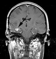
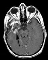
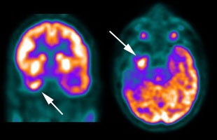
CNS recurrent glioma: The patient below had a history of a right temporal lobe glioma. The lesion had been treated with surgery and radiation. A follow-up MR exam demonstrated areas of enhancement in the right temporal lobe on post-gadolinium images (black arrows). MR spectroscopy was inconclusive. The FDG PET exam demonstrated a hypermetabolic focus in the area of MR signal abnormality consistent with recurrent glioma (white arrows). |
|
|
In the post-treatment setting, FDG PET imaging can provide
additional information which can greatly aid in patient
management. One added benefit of FDG PET imaging is in selection
of the best site for biopsy of a focal lesion [9]. Biopsy can be
directed at the most metabolically active site to enhance
diagnostic yield. FDG imaging also better defines sites of
residual tumor (when compared to MR imaging) which can
subsequently be targeted for escalated radiation therapy [12].
Findings on the FDG scan can result in a change in management in
up to 39% of patients being restaged [41].
|
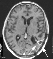
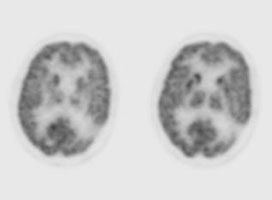
CNS radiation necrosis: The patient below had a history of a solitary brain metastasis from non-small cell lung cancer. The lesion had been resected and the patient had received radiation therapy to the area. A follow-up MR exam revealed a ring enhancing region in the left parietal-temporal area on post-gadolinium enhanced images (white arrows). A FDG PET exam revealed no tracer uptake consistent with post-surgical and post radiation change. |
|
|
MR imaging for radiation necrosis:
Hydrogen-1 (1H) MR spectroscopy can also be performed
to evaluate for radiation necrosis versus tumor recurrence [44]. A
diagnostic marker for gliomas is increased levels of
choline-containing compounds, comprising choline, phosphocholine,
and glycerophosphocholine, caused by over-expression or activation
of enzymes in choline metabolism and increased cell turnover [62].
Increased total choline levels correlate with elevated cell
proliferation rates and progress towards higher grades of
malignancies [62]. The presence of these metabolites are
detectable by MR spectroscopy [62]. The choline-creatinine ratio
and choline-N-acetyl aspartate ratio are significantly higher in
recurrent tumor and in radiation necrosis [44]. A combined
diagnostic threshold of a choline-creatine ratio greater than 1.11
and a choline-N-acetyl aspartate ratio greater than 1.17 has a
sensitivity of 89% and a specificity of 83% for the identification
of tumor [44]. An elevated lipid-lactate peak and a generalized
decrease in other metabolite levels suggests radiation necrosis
[44].
REFERENCES:
(1) J Nucl Med 1994; Ishizu K, et al.
Effects of hyperglycemia on FDG uptake in human brain and glioma.
35 (7): 1104-09
(2) Cancer 1988, 62:1074-78
(3) J Nucl Med 1994; Dethy S, et al. Carbon-11-methionine and fluorine-18-FDG PET study in brain hematoma. 35 (7) 1162-66
(4) J Nucl Med 1995; Ogawa T, et al. Carbon-11-methionine PET evaluation of intracerebral hematoma: Distinguishing neoplastic from non-neoplastic hematoma. 36 (12): 2175-79
(5) J Comput Assist Tomogr 1993; Ishii K, et al. High L-methyl-[11C] methionine uptake in brain abscess: A PET study. 17: 660-1 (No abstract available)
(6) Acta Radiol 1991; Ogawa T, et al. Clinical value of PET with 18F-fluorodeoxyglucose and L-methyl-11C-methionine for diagnosis of recurrent brain tumor and radiation injury. 32: 197-202
(7) J Nucl Med 1995; 36 (1): 163
(8) J Nucl Med 1999; Delbeke D. Oncological applications of FDG PET imaging: Brain tumors, colorectal cancer, lymphoma, and melanoma. 40: 591-603
(9) J Nucl Med 2001; Langleben DD, Segall GM. PET in differentiation of recurrent brain tumor from radiation injury. 41: 1861-1867
(10) J Nucl Med 2001; Jager PL, et al. Radiolabeled amino acids: Basic aspects and clinical applications in oncology. 42: 432-445
(11) Radiol Clin N Am 2001; Hagge RJ, et al. Positron emission tomography. Brain tumors and lung cancer. 39: 871-881
(12) J Nucl Med 2002; Tralins KS, et al. Volumetric analysis of 18F-FDG PET in gliobastoma multiforme: prognostic information and possible role in definition of target volumes in radiation dose escalation. 43: 1667-1673
(13) J Nucl Med 2004; Henze M, et al. PET and SPECT for detection of tumor progression in irradiated low-grade astrocytoma: a receiver-operating-characteristics analysis. 45: 579-586
(14) J Nucl Med 2004; Levivier M, et al. Use of stereotactic PET images in dosimetry planning of radiosurgery for brain tumors: clinical experience and proposed classification. 45: 1146-1154
(15) J Nucl Med 2004; Pirotte B, et al. Comparison of 18F-FDG and 11C-methionine for PET-guided sterotactic brain biopsy of gliomas. 45: 1293-1298
(16) J Nucl Med 2004; Spence AM, et al. 18F-FDG PET of gliomas at delayed intervals: improved distinction between tumor and normal gray matter. 45: 1653-1659
(17) J Nucl Med 2005; Chen W, et al. Imaging proliferation in brain tumors with 18F-FLT PET: comparison with 18F-FDG. 46: 945-952
(18) J Nucl Med 2005; Love C, et al. FDG PET of infection and inflammation. 25: 1357-1368
(19) Radiol Clin N Am 2005; Hustinx R, et al. PET imaging for differentiating recurrent brain tumor from radiation necrosis. 43: 35-47
(20) J Nucl Med 2005; Jacobs AH, et al. 18F-fluoro-L-thymidine and 11C-methylmethionine as markers of increased transport and proliferation in brain tumors. 46: 1948-1958
(21) J Nucl Med 2006; Popperl G, et al. Analysis of 18F-FET PET for grading of recurrent gliomas: is evaluation of uptake kinetics superior to standard methods. 47: 393-403
(22) J Nucl Med 2006; Cher LM, et al. Correlation of hypoxic cell fraction and angiogenesis with glucose metabolic rate in gliomas using 18F-fluoromisonidazole, 18F-FDG PET, and immunohistochemical studies. 47: 410-418
(23) J Nucl Med 2006; Floeth FW, et al. 18F-FET PET differentiation of ring-enhancing brain lesions. 47: 776-782
(24) J Nucl Med 2006; Chen W, et al. 18F-FDOPA PET imaging of brain tumors: comparison study with 18F-FDG PET and evaluation of diagnostic accuracy. 47: 904-911
(25) J Nucl Med 2006; Muzi M, et al. Kinetic analysis of 3'-deoxy-3'-18F-fluorothymidine in patients with gliomas. 47: 1612-1621
(26) J Nucl Med 2007; Floeth FW, et al. Prognostic value of O-(2-18F-fluoroethyl)-L-tyrosine PET and MRI in low-grade glioma. 48: 519-527
(27) J Nucl Med 2007; Rutten I, et al. PET/CT of skull base meningiomas using 2-18F-fluoro-L-tyrosine: initial report, 48: 720-725
(28) J Nucl Med 2007; Wyss MT, et al. Spatial heterogeneity of low-grade gliomas at the capillary level: a PET study on tumor blood flow and amino acid uptake. 48: 1047-1052
(29) J Nucl Med 2007; Chen W. Clinical applications of PET in brain tumors. 48: 1468-1481
(30) J Nucl Med 2007; Schiepers C, et al. 18F-FDOPA kinetics in brain tumors. 48: 1651-1661
(31) Radiology 2007; Hammoud DA, et al. Molecular neuroimaging: from conventional to emerging technologies. 245: 21-42
(32) J Nucl Med 2007; Salber D, et al. Differential uptake of O-(2-18F-fluoroethyl)-L-tyrosine, L-3H-methionine, and 3H-deoxyglucose in brain abscesses. 48: 2056-2062
(33) J Nucl Med 2008; Zhao S, et al. Usefullness of 11C-methionine for differentiating tumors from granulomas in experimental rat models: a comparison with 18F-FDG and 18F-FLT. 49: 135-141
(34) J Nucl Med 2008; Terakawa Y, et al. Diagnostic accuracy of 11C-methionine PET for differentiation of recurrent brain tumors from radiation necrosis after radiotherapy. 49: 694-699
(35) J Nucl Med 2009; Ullrich RT, et al. Methyl-L- 11C-methionine PET as a diagnostic marker for malignant progression in patients with glioma. 50: 1962-1968
(36) J Nucl Med 2011; Nagata T, et al. Examination of 11C-methionine metabolism by the standardized uptake value in the normal brain of children. 52: 201-205
(37) J Nucl Med 2011; Deng H, et al. S-11C-methionine-L-cysteine:
a new amino acit PET tracer for cancer imaging. 52: 287-293
(38) J Nucl Med 2011; Heiss WD, et al. Multimodality assessment
of brain tumors and tumor recurrence. 52: 1585-1600
(39) J Nucl Med 2012; Schwarzenberg J, et al. 3'-deoxy-3'- 18F-fluorothymidine
PET
and
MRI
for
early
survival
predictions
in
patients
with
recurrent
malignant
glioma
treated with bevacizumab. 53: 29-36
(40) J Nucl Med 2012; unet V, et al. Peformance of 18F-fluoro-ethyl-tyrosine
(18F-FET) PET for the differential diagnosis of primary
brain tumor: a systematic review and metaanalysis. 53: 207-214
(41) J Nucl Med 2012; Walter F, et al. Impact of
3,4-Dihydroxy-6-18F-Fluoro-L-Phenylalanine PET/CT on Managing
Patients with Brain Tumors: The Referring Physician's Perspective.
53: 393-398
(42) J Nucl Med 2012; Galldiks N, et al. Assessment of treatment
response in patients with glioblastoma using O-(2-18F-fluoroethyl)-L-tyrosine
PET in comparison to MRI. 53: 1048-1057
(43) J Nucl Med 2012; Galldiks N, et al. Role of
O-(2-18F-Fluoroethyl)-L-Tyrosine PET for Differentiation of Local
Recurrent Brain Metastasis from Radiation Necrosis. 53: 1367-1374
(44) Radiographics 2012; Shah R, et al. Radiation necrosis in the
brain: imaging features and differentiation from tumor recurrence.
32: 1343-1359
(45) J Nucl Med 2012; Singhal T, et al. 11C-methionine PET for grading and prognostication in gliomas: a comparison study with 18F-FDG PET and contrast enhanced MRI. 53: 1709-1715
(46) J Nucl Med 2012; Idema AJS, et al. 3'-deoxy-3'- 18F-fluorothymidine
PET-derived
proliferative
volume
predicts
overall
survival in high-grade glioma patients. 53: 1904-1910
(47) J Nucl Med 2012; Yamamoto Y, et al. Correlation of 18F-FLT
uptake
with
tumor
grade
and Ki-67 immunohistochemistry in patients with newly diagnosed
recurrent gliomas. 53: 1911-1915
(48) J Nucl Med 2013; Kasenda B, et al. 18F-FDG PET
is an independent outcome predictor in primary central nerous
system lymphoma. 54: 184-191
(49) J Nucl Med 2013; Rapp M, et al. Diagnostic performance of 18F-FET PET in newly diagnosed cerebral lesions suggestive of glioma. 54: 229-235\
(50) J Nucl Med 2013; Tehrani OS, Shields AF. PET imaging of
proliferation with pyrimidines. 54: 903-912
(51) J Nucl med 2013; Heinzel A, et al. The Use of
O-(2-18F-Fluoroethyl)-L-Tyrosine PET for Treatment Management of
Bevacizumab and Irinotecan in Patients with Recurrent High-Grade
Glioma: A Cost-Effectiveness Analysis. 54: 1217-1222
(52) J Nucl Med 2014; Lizarraga KJ, et al. 18F-FDOPA
PET for differentiating recurrent or progressive brain metastatic
tumors from late or delayed radiation injury after radiation
treatment. 55: 30-36
(53) J Nucl Med 2014; Jansen NL, et al. Dynamic 18F-FET
PET in newly diagnosed astrocytic low-grade glioma identified
high-risk patients. 55: 198-203
(54) Radiology 2014; Ellingson BM, et al. Recurrent glioblastoma
treated with Bevacizumab: contrast-enhanced T1-weighted
subtraction maps improve tumor delineation and aid prediction of
survival inb a multicenter clinical trial. 271: 200-210
(55) J Nucl Med 2014; Heiss WD. Clinical
impact of amino acid PET in gliomas. 55: 1219-1220
(56) J Nucl Med 2015; Jansen NL, et al.
Prognostic significance of dynamic 18F-FET
PET in newly diagnosed astrocystic high-grade glioma. 56: 9-15
(57) J Nucl Med 2015; Dunkl V, et al. The usefullness of dynamic
o-(2-18F-fluoroethyl)-L-tyrosine PET in the clinical
evaluation of brain tumors in children. 56: 88-92
(58) J Nucl Med 2015; Uslu L, et al. Value of 18F-FDG
PPET and PET/CT for evaluaiton of pediatric malignancies. 56:
274-286
(59) J Nucl Med 2015; Fink JR, et al. Multimodality brain tumor
imaging: MR imaging, PET, and PET/MR imaging. 56: 1554-1561
(60) J Nucl Med 2016; Unterrainer M, et al. Serial 18F-FET
PET imaging of primary 18F-FET-negative glioma: does
it make sense? 57: 1177-1182
(61) J Nucl Med 2017; Lodge MA, et al. Repeatability of 18F-FLT
PET in a multicenter study of patients with high-grade glioma. 58:
393-398
(62) J Nucl Med 2018; Mauler J, et al. Spatial relationship of
glioma volume derived from 18F-FET PET and volumetric
MR spectroscopy imaging: a hybrid PET/MRI study. 59: 603-609
(63) AJR 2018; George E, et al. Voxel-wise analysis of
fluoroethyltyrosine PET and MRI assessment of recurrent
glioblastoma during antiangiogenic therapy. 211: 1342-1347
(64) J Nucl Med 2020; de Zwart PL, et al. Diagnostic accuracy of
PET tracers for the differentiation of tumor progression from
treatment-related changes in high-grade glioma: a systemic review
and metaanalysis. 61: 498-504
(65) J Nucl Med 2020; Maurer GD, et al. 18F-FET PET
imaging in differentiating glioma progression from
treatment-related changes: a single-center experience. 61: 505-511
(66) J Nucl Med 2020; Filss CP, et al. Flare phenomenon in O-(2-
18F-fluoroethyl)-L-tyrosine PET aterresection of
glioma. 61: 1294-1299
(67) AJR 2020; Ozutemiz C, et al. The role of dual-phase FDG
PET/CT in the diagnosis and follow-up of brain tumors. 215:
985-996
(68) AJR 2020; Sharma A, Kumar R. Metabolic imaging of brain
tumor recurrence. 215: 1199-1207
