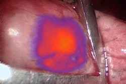
A new tumor-specific fluorescent imaging agent can help surgeons find and remove additional malignant tissue in ovarian cancer patients during surgery, according to a study published online in Clinical Cancer Research.
Lead author Dr. Alexander Vahrmeijer, PhD, of Leiden University Medical Center in the Netherlands, and colleagues developed the agent, OTL38, which combines fluorescent dye and a folate facsimile. OTL38 binds to folate receptor alpha (FRα), which is expressed in more than 90% of ovarian cancers. To image the agent, the group used near-infrared light (Clin Cancer Res, June 15, 2016).
First, the researchers conducted a randomized, double-blind, placebo-controlled clinical trial to assess the tolerability and pharmacokinetics of OTL38 in 30 healthy volunteers. Then they tested the agent in 12 patients with ovarian cancer, assessing whether OTL38-guided surgery resulted in the detection of more cancers.
The researchers found that OTL38 did accumulate in FRα-positive tumors and metastases, allowing surgeons to remove an additional 29% of malignant lesions that could not be visually identified or palpated.
"The main advantage of near-infrared light is that it can penetrate tissue in the order of centimeters, allowing the surgeon to visualize tumors underneath the tissue surface that can be detected using a dedicated imaging system," Vahrmeijer said in a statement released by the American Association for Cancer Research (AACR).


