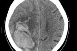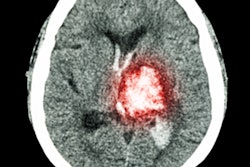
U.K. researchers have identified five factors -- including contrast leakage on CT scans -- that may indicate a higher risk of continued bleeding in the brain of stroke patients. Checking for these factors may help improve survival, according to an article published online on August 14 in Lancet Neurology.
Considered one of the most dangerous forms of stroke, intracerebral hemorrhage (ICH) leads to death in more than half of all cases and permanent brain damage in roughly 80% of the remaining survivors, noted co-first author Rustam Al-Shahi Salman, PhD, and colleagues from the University of Edinburgh.
Clinicians traditionally diagnose ICH with a standard CT or MRI exam, the authors added. However, effectively monitoring and treating patients with the condition is challenging because bleeding continues in the brains of these patients often long after the initial stroke. Identifying the risk factors that trigger this extended bleeding has been a research priority in the community.
Salman and colleagues reviewed all studies concerning intracerebral hemorrhage that were in the Ovid Medline database and published between January 1970 and December 2015. Among thousands of articles, they honed in on 36 studies that provided data on 5,435 patients.
Examining these cases, the group identified five key risk factors that independently had a statistically significant association with continued bleeding in the brain after having a stroke:
- Longer time from symptom onset to baseline CT or MRI
- Greater amount of bleeding visible on baseline CT or MRI
- Patient use of antiplatelet medication such as aspirin
- Patient use of anticoagulant medication such as warfarin
- Presence of the "spot sign," i.e., contrast leakage, on CT angiography
| Effect of risk factors on brain bleeding in stroke patients* | |
| Factor | Odds ratio for continued brain bleeding |
| Shorter time from symptom onset to baseline CT or MRI | 0.50 |
| Patient use of antiplatelet medication such as aspirin | 1.68 |
| Patient use of anticoagulant medication such as warfarin | 3.48 |
| Presence of the "spot sign," i.e., contrast leakage, on CT angiography | 4.46 |
| Greater amount of bleeding visible on baseline CT or MRI | 7.18 |
Decreasing the time between stroke onset and baseline CT or MRI from 5.1 hours to 1.5 hours resulted in an odds ratio of 0.5, suggesting that earlier imaging correlated with about half the likelihood of continued brain bleeding. An increase in the amount of bleeding seen on initial CT or MRI from 6 mL to 33 mL led to a more than sevenfold increase in the risk of continued bleeding.
The risk of continued bleeding was 1.68 times greater in patients who were taking antiplatelet medication and 3.48 times greater in those taking anticoagulant medication. Finally, the presence of the CT angiography spot sign increased the risk more than fourfold.
To further confirm these factors, the researchers used them to develop a risk prediction model. They found that the model had good calibration and could be useful in clinical practice for quickly estimating the risk of continued intracerebral hemorrhage.


