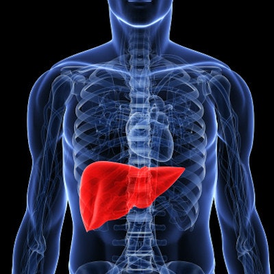
Deep-learning software can accurately stage liver fibrosis on contrast-enhanced CT studies, according to an article published online September 4 in Radiology. In a new study from South Korea, the software outperformed four radiologists as well as two serum fibrosis markers calculated from laboratory tests.
Researchers led by Kyu Jin Choi of Hanyang University and Dr. Jong Keon Jang of the University of Ulsan College of Medicine developed a deep-learning system based on two separate algorithms: one for segmenting the liver and another that stages liver fibrosis based on segmented liver data. In testing, the system yielded higher diagnostic performance than subjective evaluation by four radiologists, as well as the AST to Platelet Ratio Index (APRI) and the Fibrosis-4 index.
"The [deep-learning system] allows for highly accurate assessment of liver fibrosis by using portal venous phase CT images of the liver," the authors wrote. "Because of the widespread availability of CT and liver CT imaging, the [deep-learning system] is a promising and widely applicable method for assessment of liver fibrosis."
The researchers trained the liver segmentation algorithm using liver outlines drawn by an experienced radiologist on portal venous phase CT images from 50 randomly selected patients in a development dataset of 7,461 CT exams. The liver fibrosis staging algorithm was trained on the entire development dataset. They then validated the system clinically with three independent datasets including 891 patients with a confirmed diagnosis of liver fibrosis.
Diagnostic performance was calculated using the area under the receiver operating characteristic curve (AUROC), as well as the Obuchowski index -- a weighted average of AUROC values that estimates the probability that a test will correctly rank two randomly chosen patients with different stages of fibrosis. The results were then compared with serum fibrosis markers and the findings of four radiologists who had independently reviewed and staged the CT images; radiologists 1 to 3 were academic abdominal radiologists, while radiologist 4 was a trainee abdominal radiology fellow.
| Diagnostic performance of AI algorithm for liver fibrosis staging | |||||||
| Radiologist | APRI | Fibrosis-4 index | Deep-learning algorithm | ||||
| 1 | 2 | 3 | 4 | ||||
| Obuchowski index | 0.77 | 0.78 | 0.81 | 0.74 | 0.71 | 0.76 | 0.94 |
The difference in staging performance between the deep-learning algorithm and the radiologists, APRI, and fibrosis-4 index was statistically significant (p < 0.01). The researchers pointed out that radiologists who interpret liver CT images do not routinely stage liver fibrosis in clinical practice, "although the presence of chronic liver disease or cirrhosis is sometimes suggested on the basis of typical imaging findings."
While the test dataset included data from various scanners and CT techniques, the researchers also noted that there has been continual progress in CT methods, including new image reconstruction algorithms for lowering radiation dose.
"Therefore, the applicability of the [deep-learning system] when novel CT techniques are used should also be evaluated in future studies," they wrote. "Finally, further research would be required to evaluate the clinical benefits of the [deep-learning system] in predicting the prognosis and helping to guide management of patients with chronic liver disease."


