CT scans helped clinicians identify and remove a 3-cm needle from a patient who swallowed it, likely during a dental procedure, according to a recent case report published in the April issue of the Journal of Dental Anesthesia and Pain Medicine.
This is believed to be the first reported case of an ingested dental needle causing neck pain, the authors wrote.
"Since human error is inevitable, we must create systems and follow safety measures to reduce the incidence of [foreign bodies] and prioritize patient safety," wrote the group, led by Dr. Hassen Mohammed of Hamad Medical Corp. in Doha, Qatar (J Dent Anesth Pain Med, April 2020, Vol. 20:2, pp. 83-87).
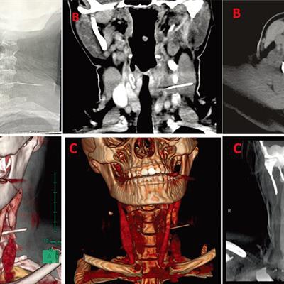
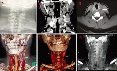 A: An x-ray showed a long, thin, and impinged foreign body. B: CT confirmed that the body was metallic and 3 cm in length. C: 3D reconstructed images were used for planning the removal of the needle. All images courtesy of Dr. Hassen Mohammed et al. Licensed under CC BY-NC 4.0.
Rare but not impossible
Disposable stainless-steel needles have proved to be a safe and effective method for dental procedures such as root canal washes, and complications are rare. However, in some cases, dental needle breakages have occurred when patients bit needles or clinicians used incorrect injection techniques or inadequate preventative measures. Sometimes, patients have swallowed these broken needles during procedures.
In this case, a 29-year-old woman likely unknowingly swallowed a needle during a dental procedure. She had some throat pain after the procedure, and a few days later, she began experiencing neck pain, swelling, and a foreign-body sensation.
She told clinicians that she began feeling pain after eating duck and assumed that she had swallowed a bone. She went to a primary healthcare center, where she was prescribed painkillers and antibiotics for pharyngitis. No imaging was performed during that visit.
The pain persisted for 12 days, so she returned to the center. Once there, she underwent a soft-tissue neck x-ray. The x-ray detected a foreign object that turned out to be a dental needle, and the patient was referred immediately to the ear, nose, and throat department.
After a thorough exam and a flexible fiber-optic nasopharyngolaryngoscopy didn't reveal the foreign body in her upper aerodigestive tract, the clinicians saw a tender, nonfluctuant, diffuse swelling of about 2 x 2 cm on the left side of her neck. The x-ray showed a long, thin, smooth, and impinged foreign body, lying lateral to the esophagus. CT showed that something metallic and 3 cm in length had pierced through the woman's left internal jugular vein and the left sternocleidomastoid muscle. Doctors used 3D reconstruction and virtual endoscopy images to localize the needle and plan for the surgery, the authors wrote.
Probably how it happened
After surgically removing the needle, having further discussions with the patient, and showing her the needle that was removed, the clinicians concluded that she probably ingested the needle during a dental procedure that occurred on the same day she had eaten the duck. She didn't remember much about the procedure because she had undergone general anesthesia for the treatment.
Furthermore, the authors found this to be the most logical explanation after they attempted a simulation to compare needle sizes and structure. They used a 23-gauge needle and bent it as if it were going to be used for a dental procedure. The needle was almost identical to the one removed from the woman's neck.
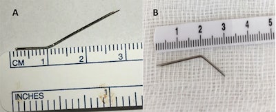 A: The needle removed from the woman's neck. B: The simulated needle, which shows they are almost identical.
The takeaway
Virtual endoscopy and 3D reconstruction should be used to aid the diagnosis of airway pathology because they help clarify the location and characteristics of foreign bodies. Also, a multidisciplinary team of surgeons, radiologists, and anesthesiologists should be consulted when difficult airway management cases occur. Finally, dentists should use larger needles when available and take extra care when injecting patients, they wrote.
"Clinicians performing intraoral procedures should also use larger-gauge needles when possible, as smaller needles are more susceptible to accidental breakages," the authors wrote.


