Chest x-rays are effective for the early detection of COVID-19, especially in older patients, according to a study presented at the 2021 virtual annual meeting of the American Roentgen Ray Society (ARRS).
Researchers found that chest radiography was more effective in diagnosing COVID-19 in people older than 50 years. In addition, there is a specific pattern of abnormalities on chest x-rays that can be used to diagnose the disease, they reported. The findings add to the body of knowledge regarding the use of radiography during the pandemic.
"Compared with chest CT, there is a relative paucity of data regarding the role of chest radiography in the diagnosis of COVID-19," said Dr. Mae Igi of the Louisiana State University Health Sciences Center in New Orleans.
In this retrospective study, Igi and colleagues looked at the utility of a strict pattern of findings at chest x-ray for the diagnosis of COVID-19, particularly during early onset of symptoms in patients older and younger than 50.
The study involved individuals presenting to the emergency room during the first phase of the COVID-19 outbreak in March 2020. Sixty patients found to be positive for COVID-19 on reverse transcription polymerase chain reaction (RT-PCR) tests were included; patients had chest x-rays within one week of reported symptoms.
The patients were split into two groups: those older than 50 years and those younger than 50. The age range was 52 to 88 years in the older group and 19 to 48 years in the younger group.
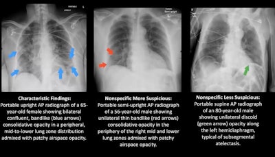
Two board-certified radiologists who were blinded to the RT-PCR results assessed each of the 60 chest x-rays in consensus and classified the images into one of three patterns: characteristic for COVID-19, nonspecific for COVID-19, and negative. The nonspecific patterns were further broken down into "more suspicious" or "less suspicious" for COVID-19, Igi explained.
In the older group, 93% demonstrated an abnormal chest x-ray early in the course of COVID-19 disease. In comparison, 53% of the younger group demonstrated an abnormal chest x-ray early in the course of the disease.
The relationship between age and chest x-ray findings was determined to be statistically significant (p = 0.00039). The relationship was confirmed with a Fisher exact test.
 Image courtesy of Dr. Mae Igi.
"COVID-19-positive patients over 50 show earlier, more characteristic patterns of statistically significant [chest x-ray] changes than younger patients, suggesting that [chest x-ray] is useful in the early diagnosis of the disease," Igi concluded.
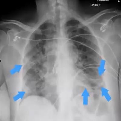
What are characteristic findings on chest x-ray that are telltale signs of COVID-19? They can include the presence of bilateral patchy or confluent bandlike ground-glass opacity, or consolidation in a peripheral or middle to lower lung zone.
On the other hand, abnormal findings that are less suspicious for COVID-19 can include pulmonary edema, atelectasis, and interstitial changes.
In another study presented during the session, Dr. Emily Tsai, a thoracic radiologist at Stanford University, encouraged the use of a technique called "through glass" chest x-ray for diagnosing COVID-19. The technique, initially developed by University of Washington researchers in Seattle during the 2014 Ebola outbreak, involves performing radiography through the glass of isolation room doors with portable machines.
Tsai said they have implemented the technique at Stanford and observed no significant reduction in the quality of imaging when compared to regular chest x-rays. The technique reduces exposure to front-line workers and conserves personal protective equipment, according to Tsai.
"The COVID-19 pandemic has forced us to consider carefully how we use imaging, and it has required us to adapt quickly as we continually learn more about this evolving disease," she concluded.


