Monday, November 26 | 11:00 a.m.-11:10 a.m. | SSC09-04 | Room S504CD
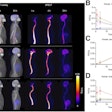
F-18 sodium fluoride (NaF) PET/CT detects more skeletal lesions than technetium-99m (Tc-99m) methylene diphosphonate (MDP) SPECT in patients newly diagnosed with breast cancer, according to researchers from Tata Memorial Centre in Mumbai, India.The study was led by Dr. Venkatesh Rangarajan, head of the department of nuclear medicine and molecular imaging. The group evaluated 30 patients with newly diagnosed breast cancer who underwent NaF-PET scans along with whole-body contrast-enhanced CT as part of the combined PET/CT study. An MDP bone scan also was performed within a week of the PET/CT study.
"In locally advanced breast cancer, the current [National Comprehensive Cancer Network (NCCN)] protocol recommends an MDP bone scan and contrast-enhanced CT for the staging," Rangarajan said. "In this scenario, knowing the incremental value of NaF-PET/CT over bone scans with higher accuracy, we integrated contrast-enhanced CT with NaF-PET as an integrated protocol."
Lesions in the MDP bone scan and NaF-PET/CT bone scan were compared and classified as benign, malignant, or equivocal.
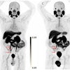
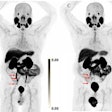
In the analysis, MDP bone scans detected 84 lesions: 20 were benign, 44 were metastatic, and 20 were equivocal. NaF-PET/CT detected 162 lesions: 113 were metastatic, 49 were benign, and none were classified as equivocal.
NaF-PET/CT was positive in five patients who were negative with MDP, according to the researchers.
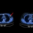
Contrast-enhanced CT detected metastases in 10 of the 30 patients, which included metastases in five lungs, five livers, three axillary nodes, and one each in the brain and adrenal area. NaF-PET/CT helped to detect lesions and changed treatment management in two patients, who initially appeared normal on the MDP bone scan.
NaF-PET/CT is "very patient-friendly and provides comprehensive staging along the recommendations of NCCN," Rangarajan said.


