Sunday, November 27 | 11:35 a.m.-11:45 a.m. | SSA16-06 | Room S505AB
Even though PET imaging with fluorocholine or sodium fluoride provides different information on patients with prostate cancer, either PET option appears to be better than CT for assessing the extent of active metastatic skeletal disease."The major finding is that CT cannot be used for assessment of bone therapy response in the skeletal metastases of prostate cancer, external-beam radiation therapy of oligometastatic disease, chemotherapy of castration-resistant metastatic disease, or palliative bone pain radionuclide treatments, and in general in the follow-up of skeletal disease in prostate cancer," said Dr. Kalevi Kairemo, PhD, from MD Anderson Cancer Center. "Therefore, PET should be applied in these conditions."
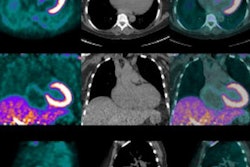
Kairemo and colleagues reviewed 12 patients with advanced skeletal prostate cancer of more than five lesions who were scanned on consecutive days by PET/CT both with fluorocholine and sodium fluoride. Bone regions as seen in CT were used to coregister the two PET/CT scans.
The researchers defined volumes of interest for fluorocholine and sodium fluoride PET uptake and overlapped the results with CT-based skeletal and sclerotic volumes of interest. They found no statistical difference for sclerosis on CT between patients with metastases and those without metastases.
By comparison, PET with fluorocholine or sodium fluoride significantly differentiated between variables such as total bone demineralization activity, total lesion glycolysis, and skeletal volume in prostate cancer patients and control subjects with no metastases.
"CT demonstrates only sites where there sometimes has been metastatic activity," said Kairemo, who also is a professor of molecular radiotherapy and nuclear medicine at the Docrates Cancer Center in Helsinki. "Both PET methods performed within one day in this study gave very different information from each other and from CT and complementary information about the nature of the skeletal disease in these patients."


