Researchers at Washington University School of Medicine in St. Louis have developed a chemical compound that detects amyloid deposits on PET scans better than currently approved imaging agents, according to a paper published November 2 in Scientific Reports, one of the Nature journals.
The compound is known as fluselenamyl. When a radioactive atom is incorporated into the compound, the atom's location can be monitored through PET in a living brain and potentially identify the signs of early-stage Alzheimer's disease or monitor treatment response.
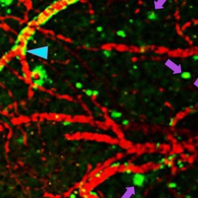
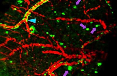
 In the image above, fluselenamyl has passed from the bloodstream of a living mouse into its brain, where it is detected by a PET scan. Arrows indicate clumps of beta amyloid. Image credit: Ping Yan and Jin-Moo Lee.
Senior author Vijay Sharma, PhD, a professor of radiology, neurology, and biomedical engineering at Washington University, said fluselenamyl is both more sensitive and likely to be more specific than current agents, and it reduced the number of false negatives in this study.
Using human beta-amyloid proteins, the researchers found that fluselenamyl bound to proteins two to 10 times better than three imaging agents currently cleared by the U.S. Food and Drug Administration (FDA) for detecting beta amyloid.
The group also used the compound to stain brain slices from people who had died of Alzheimer's disease, along with a control cohort of people of similar ages who had died of other causes. Fluselenamyl was able to identify plaques in the brain slices of the Alzheimer's patients but not in the control group.
The researchers have submitted an application to the U.S. National Institutes of Health (NIH) for a phase 0 trial to determine if fluselenamyl is safe for use in humans and behaves in the human body the same way it behaves in mice.


