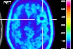
When glioma results are inconclusive on MRI, clinicians should consider fluoroethyl-tyrosine (FET)-PET for more definitive results and to differentiate between tumor progression and treatment improvement, according to a study published online September 13 in the Journal of Nuclear Medicine.
German and Dutch researchers found that the FET-PET achieved sensitivity, specificity, and accuracy of 70% or greater and helped solve mysteries left by MRI.
"Diagnosis and treatment of brain tumors are strongly linked to imaging, especially MRI, techniques, as histological confirmation often cannot be realized easily and without substantial risk," wrote the authors, led by Dr. Gabriele Maurer from Goethe University Hospital in Frankfurt. "FET-PET is not a standard method for the assessment of in glioma, but may be more accurate than conventional MRI and helpful in complex or challenging cases."
Among the challenges for clinicians is differentiating between tumor progression and treatment-related changes in glioma cases. The authors referred to this "frequent problem" and "pseudoprogression," in which a contrast-enhanced area enlarges more than 25%, even when there is no tumor, or when new lesions occur during or after therapy. These abnormalities mimic tumor progression on MRI scans within the first three months after chemoradiation.
"In our neuro-oncology department, we recommended FET-PET imaging when conventional MRI and clinical assessment left some ambiguity as to whether tumor progression or sequelae of therapy prevailed," they noted. "We here outline our experiences and focus on the diagnostic performance of additional 18F-FET PET scans in clinical routine."
This retrospective study included 127 patients (median age, 50 years ± 12 years; range, 20-78 years) who were referred for evaluation of tumor progression and treatment-related changes. Of those subjects, 125 people (98%) previously were diagnosed with grade II-IV gliomas, while the two other patients were treated for suspected glioma without prior biopsy. FET-PET imaging was performed on either a standalone scanner (Ecat Exact HR+, Siemens Healthineers) or PET/MRI system (BrainPET, Siemens) and began 0 to 50 minutes after injection of 3 MBq/kg based on body weight.
Maurer and colleagues were able to diagnose tumor progression in 94 patients (74%) and treatment-related changes in 33 cases (26%). Receiver operating characteristic (ROC) analysis showed an optimal FET maximum tumor-to-background ratio (TBRmax) cutoff value of 1.9 for differentiating between tumor progression and treatment-related changes. That benchmark resulted in sensitivity of 70%, specificity of 71%, and accuracy of 70%.
The researchers also delved into how tumor characteristics, such as isocitrate dehydrogenase (IDH) mutations, affected FET-PET accuracy. In these cases, FET-PET diagnoses were incorrect in 17 patients (33%) with 51 IDH-mutant tumors (8 false positive and 9 false negatives).
This latter finding "could be helpful when considering FET-PET as an additional diagnostic tool," wrote Maurer and colleagues, suggesting that it might have been missed in previous studies that lacked molecular markers, had smaller collectives, or had a minor fraction of patients with treatment-related changes. "It is certainly worth further investigation and should be verified in a larger number of patients."


