PET imaging has revealed reduced FDG uptake in key brain regions associated with early- and late-stage Alzheimer's disease among people with Down syndrome. The findings could indicate the degree of brain atrophy in individuals, as well as one reason why this patient population is susceptible to dementia.
"As we collect more data and more participants become amyloid-positive, tau-positive, and neurodegeneration-positive, we can pinpoint which regions show the greatest atrophy and FDG hypometabolism," said lead author Matthew Zammit, PET research assistant at the University of Wisconsin-Madison. Zammit presented the findings this month at the Society of Nuclear Medicine and Molecular Imaging (SNMMI) virtual annual meeting.
Adults with Down syndrome are predisposed to Alzheimer's disease due the triplication of chromosome 21, which results in the increased production of amyloid protein and an early presence of beta-amyloid plaques in the brain's striatum, which contains the caudate and is responsible for motor coordination. To determine the severity of Alzheimer's, clinicians have turned to an amyloid/tau/neurodegeneration (ATN) framework that includes beta-amyloid-PET, tau-PET, and FDG-PET/MRI, respectively.
"Within this [ATN] framework, it is speculated that FDG-PET and MRI can classify neurodegeneration," Zammit told the attendees at the virtual SNMMI session. "However, previous studies with FDG and Down syndrome have been limited by small sample sizes, so the usefulness of FDG-PET within the ATN framework has yet to be evaluated."
Thus the goal for Zammit's study was to analyze beta-amyloid deposition using PET with carbon-11-labeled Pittsburgh Compound B (PiB) and compare it with glucose metabolism of FDG-PET among people with Down syndrome while measuring their progression to Alzheimer's disease.
The researchers recruited 90 adults (age 38 ± 8.3 years) with Down syndrome from the Alzheimer's Biomarker Consortium-Down Syndrome (ABC-DS), which contains beta-amyloid PET data on more than 300 adults with Down syndrome spanning 10 years.
"We do have a very young cohort, but this cohort is in that 'sweet spot' of when they are becoming amyloid-positive," Zammit explained. "We were able to bridge the connection to when they become amyloid-positive and how many years until they experience neurodegeneration. The average age of dementia onset in Down syndrome is about 55."
Subjects underwent PiB- and FDG-PET scans, along with T1-weighted MRI scans for anatomical reference and brain volume measurements. Standardized uptake value ratios (SUVr) were calculated using PiB and FDG uptake in the cerebellum, which served as the reference region. Based on those results, an SUVr cutoff of 20% indicated beta-amyloid positivity.
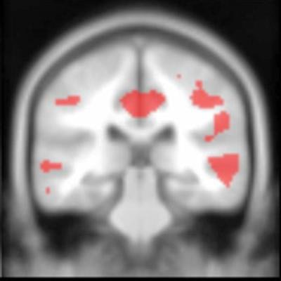
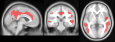
The researchers found significant levels of reduced FDG uptake (hypometabolism) with increases of beta amyloid, which could indicate brain atrophy. The most significant declines in FDG uptake (p < 0.05) were observed in the perinatal cortex, precuneus, and posterior cingulate, which typically are associated with early-stage Alzheimer's.
Conversely, the authors noted significant FDG uptake (hypermetabolism) with greater beta-amyloid load, with the most significant increases (p < 0.05) in the putamen and thalamus. All of the changes in FDG were driven entirely by subjects who were beta-amyloid-positive.
 PET images show significant negative associations between PiB and FDG in the cingulate, temporal cortex, parietal cortex and precuneus. Image set courtesy of Journal of Nuclear Medicine (JNM, May 1, 2020; vol. 61: Supplement 1).
As for the striatum and early signs of Alzheimer's disease in Down syndrome subjects, FDG reductions were observed in the caudate but were not statistically significant (p = 0.53). The FDG decrease "strongly correlated with ventricle volume," Zammit added, "suggesting that the FDG reduction may be influenced by ventricular enlargement that is common in Down syndrome with Alzheimer's disease."
Given the measurable declines in FDG uptake related to Alzheimer's disease and Down syndrome, the findings suggest that "FDG is a strong marker for neurodegeneration in the ATN framework in this population," Zammit concluded. However, while the "early-stage [beta-amyloid] accumulation in the striatum can provide amyloid-positive information, this region does not inform neurodegeneration status when measured with FDG."


