Nuclear medicine physicians in France have identified a metabolic pattern on brain PET images that indicates the presence of long COVID-19 in patients, according to a study published March 23 in the European Journal of Nuclear Medicine and Molecular Imaging.
The pattern shows hypometabolism in the fronto-orbital olfactory regions of the brain and limbic and paralimbic regions, as well as the brainstem and cerebellum, the authors wrote.
"The proposed PET metabolic pattern is easily identified upon visual interpretation in clinical routine for approximately one half of patients with suspected neurological long COVID," wrote lead study author Dr. Eric Guedj, PhD, head of nuclear medicine at Marseille University Hospital.
Long COVID is defined by the persistence or recurrence of symptoms three months after an initial infection with the SARS-CoV-2 virus. Long COVID represents a serious public issue because it affects up to 15% of patients, with emerging evidence of brain impairment, according to the authors.
In recent previous work, the researchers described the hypometabolic pattern as observed on a group level on F-18 FDG-PET scans of 35 patients compared with 44 healthy patients who underwent imaging at a single institution, Timone hospital in Marseille.
"Validation of the long-COVID hypometabolic PET pattern is consequently needed at the individual and multicentric levels to easily apply it in clinical routine for nuclear physicians," they wrote.
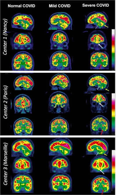
To that end, the group aimed to validate the pattern with experienced nuclear medicine physicians at three hospitals (in Nancy, Paris, and Marseille) using it to visually interpret F-18 FDG-PET imaging of suspected long-COVID patients as either normal, mildly to moderately affected, or severely affected.
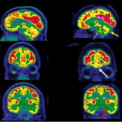
 Typical examples of brain F-18 FDG-PET images of patients with suspected long COVID in three French nuclear medicine departments, all presenting positive RT-PCR and/or serology tests. In patients with a typical severe long-COVID hypometabolic pattern, the brain areas with hypometabolism are identified with arrows: white arrows for the fronto-orbital olfactory regions, red arrows for the other limbic/paralimbic regions, grey arrows for the pons, and yellow arrows for the cerebellum. Image courtesy of the European Journal of Nuclear Medicine and Molecular Imaging.
The group analyzed F-18 FDG-PET images of 143 patients (47.4 years old ± 13.6; 98 women) referred for suspected neurological long COVID with positive reverse transcription polymerase chain reaction (RT-PCR) tests between August 2021 and October 2021.
All patients met the current definition of long COVID by the World Health Organization, with a PET scan performed at least three months after infection and while their symptoms persisted. Patients reported typical long COVID symptoms: fatigue, pain, memory and cognitive complaints, insomnia, loss of smell, loss of taste, and signs of dysautonomia (tachycardia, orthostatic intolerance, and breathlessness).
Based on the visual analysis, 53% of the PET scans were interpreted as normal, 21% as mildly to moderately affected, and 26% as severely affected according to the COVID hypometabolic pattern, the researchers found.
"This pattern can be found at an individual level and helps to objectively assess metabolic abnormalities in patients with suspected neurological long COVID," the group wrote.
Of note, the regions affected in the PET pattern are known to be related to typical symptoms of patients with long COVID, especially olfactory loss, emotional disturbances, memory impairment, motor disorders, impaired balance, and dysfunction of autonomous behaviors, they added.
Moreover, the pattern is clearly different from patients with neurodegenerative and psychiatric diseases, they wrote.
Ultimately, the exam could reassure half of long COVID patients suspected of brain impairment who have normal scans, the researchers suggested. On the other hand, it could contribute to social and medical recognition of long COVID in patients, especially when all other medical examinations fail to show an objective abnormality, they wrote.
"Nuclear neurology can be helpful in routine practice for selected patients, here on average 10.9 months from the initial infection, after clinical evaluation, to identify the possible brain impairment associated with long COVID," Guedj and colleagues concluded.


