Hybrid PET/MRI is leading the way in imaging technologies applied in the emerging field of cancer theranostics, according to a presentation at the International Society for Magnetic Resonance in Medicine (ISMRM) annual meeting.
In a June 7 plenary session, Lisa Bodei, MD, PhD, a nuclear medicine physician at Memorial Sloan Kettering Cancer Center in New York, NY, discussed how she uses PET/MRI to identify patients with neuroendocrine tumors and prostate cancer.
"PET/MRI may replace standalone MRI and PET/CT applications where the anatomic information from MRI is fundamental," she said. "And these include theranostics, particularly for neuroendocrine tumors and prostate cancer."
Theranostics is a portmanteau of "therapy" and diagnostics" and refers to the dual approach of using the same molecular ligands to both image and treat cancer. It is an emerging field in nuclear medicine, Bodei explained.
Hybrid PET/MRI, which was developed about 12 years ago, combines both PET and MRI modalities in a single scanner for near-simultaneous acquisitions. These scanners provide clinicians with detailed anatomical tumor assessments from multiparametric MRI with complementary physiologic information on tumors based on injected PET radiotracer uptake.
PET imaging radiotracers used in theranostics include gallium-68 (Ga-68) DOTATATE, while therapeutic drugs include lutetium-177 DOTATATE (Lutathera), for instance. In patients with NETs – a type of cancer that often metastasizes from primary sites to other sites in the body – PET/MRI fusion imaging using Ga-68 DOTATATE is proving essential, Bodei noted.
Specifically, she presented cases in patients with metastatic neuroendocrine tumors in the liver, bone, adrenal glands, and the brain. These lesions were detected using PET/MRI when either approach alone or PET/CT imaging was not enough, she said.
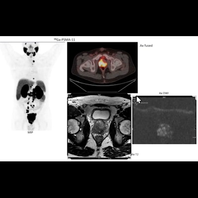
 PET/MRI imaging in a patient with prostate cancer illustrating how PET added value in fused images where cancer was difficult to detect on MRI alone. Image courtesy of Lisa Bodei, MD, PhD.
Ultimately, the shift toward the use of integrated PET/MRI in patients with neuroendocrine tumors is "inevitable," Bodei noted, as these are the two most accurate diagnostic techniques for these patients.
Similarly, in prostate cancer imaging, PET/MRI with prostate-specific membrane antigen (PSMA) radiotracers such as Ga-68 PSMA-11 is proving to be a game changer and could eventually even replace the need for invasive biopsies during initial treatment planning for patients, she said. Many of these patients are treated with Lu-177 PSMA-617 (Pluvicto).
Studies suggest that multiparametric MRI is better than PSMA-PET for localizing primary tumors (98% vs. 86%, respectively) in these settings, while PSMA-PET is better for correctly identifying prostate cancer that has metastasized, particularly to lymph nodes (60% vs. 50%), and thus combined, the technology could provide superior accuracy, she said.
"This is a new concept that is creating a lot of interest," she said. "Can we avoid a biopsy utilizing PSMA-PET or MRI, or even better, using PSMA-PET/MR?"
She cited a recent German study that showed how PET/MRI localized prostate cancer in patients without biopsies in 98% of 25 cases.
Specific limitations of PET/MRI include the length of the scans, however, which are complex.
"It is vital that we optimize the number of sequences, and we keep the duration of the scan below one hour," Bodei concluded.