Adding focused ultrasound (FUS) to MR-PET may be a feasible and cost-effective method for therapy monitoring, according to findings to be presented May 12 at the International Society for Magnetic Resonance in Medicine (ISMRM) annual meeting.
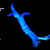
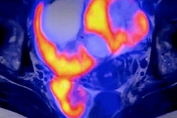
 Researchers at ISMRM highlighted the success of a trimodal MR-PET-FUS system built around a low-field MRI scanner. MR (top) and PET (bottom) images were acquired simultaneously before and after FUS heating.ISMRM
In a digital poster presentation, a team led by Teresa Guallart-Naval from the Institute for Instrumentation in Molecular Imaging (i3M) in Valencia, Spain, highlighted its trimodal MR-PET-FUS system’s performance in a phantom study, which produced clear images and achieved simultaneous PET-MR imaging while ultrasound-induced hyperthermia was applied. The researchers built the system around a low-field MRI scanner.
“This low-field hybrid technology opens new possibilities for real-time therapy monitoring and diagnostic applications, especially for settings where conventional MRI-PET systems are impractical or inaccessible,” Guallart-Naval said in her digital presentation.
Low-field MRI devices address the limitations of standard MRI scanners, such as bulky size, logistical challenges, and cost. Furthermore, low-field MRI integrates well with PET and focused ultrasound.
Guallart-Naval and colleagues highlighted that with these advantages in mind, low-field MRI could be a good starting point for hybrid imaging modalities. For the study, the team presented what it calls the first trimodal MR-PET-FUS system built around a low-field MRI scanner.
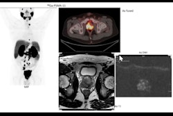
 Acquisitions show images during the FUS application. Top: a) average of 75 images acquired every minute, and b) profiles along the central vertical line of the phantom. Bottom: time evolution of c) image amplitude at the center of the phantom, d) measured voltage at center of k-space, e) measured noise, and f) signal-to-noise ratio.ISMRM
To create the system, the researchers integrated a 72-mT Halbach MRI scanner with PET and FUS technologies. They tested its performance on a cylindrical phantom containing a copper sulfate solution, gelatin, and a radioactive tracer.
While initial PET-MRI showed separation between the gelatin and copper sulfate regions, FUS heating caused the gelatin to melt and mix, causing homogenization within the phantom.
The team reported that it acquired PET-MR images while maintaining MRI operation with increased thermal noise, about three times the Johnson noise level.
“This noise increase is due to electronic interference from the PET components and did not prevent image acquisition, but it did affect image quality,” it wrote.
Also, signal and noise both increased during heating, with the former stabilizing at about 10% above the initial noise. The researchers suggested that this is likely due to sample heating. However, the signal increased by up to 100%, resulting in a better signal-to-noise ratio. They also noted that the system had minimal inter-system interference.
“Having demonstrated simultaneous MR-PET acquisition and FUS heating, this work opens the path for cost-effective, multimodal diagnostics and treatment applications,” the researchers wrote.
Check out AuntMinnie.com’s full coverage of ISMRM 2025 here.