A new PET/CT radiotracer that detects insulinomas could eliminate the need for other diagnostic procedures in a vast majority of patients, according to a study published October 17 in the Journal of Nuclear Medicine.
The finding is from a clinical trial that compared PET/CT imaging with gallium-68 (Ga-68) NODAGA-exendin-4 PET/CT with all routine imaging procedures for the localization of insulinomas.
“Because of the high sensitivity and excellent imaging quality shown here, exendin PET/CT should be considered as a potential primary diagnostic imaging modality in patients with [endogenous hyperinsulinemic hypoglycemia],” wrote lead author Marti Boss, MD, of Radboud University Medical Center in Nijmegen, the Netherlands.
Insulinomas are small neuroendocrine tumors of the pancreas that produce too much insulin. They are the most common cause of endogenous hyperinsulinemic hypoglycemia (EHH), which can lead to symptoms such as confusion, diplopia, and dizziness -- and in prolonged cases, even seizures or death, the authors explained.
Identifying these tumors for curative surgery relies on imaging, yet current routine noninvasive imaging techniques, including CT, MRI, and Ga-68 DOTA-somatostatin analog (SSA) PET/CT, have limited sensitivity, the researchers noted. Endoscopic ultrasound is highly sensitive, but invasive, they added.
In previous studies, Ga-68 NODAGA-exendin-4 has been shown to bind to a receptor on insulinomas with high affinity, and in this study, the researchers aimed to further assess its potential clinical value.
The group enrolled 69 adults with EHH from five centers in the Netherlands and France. All participants underwent Ga-68 NODAGA-exendin-4 (exendin) PET/CT and at least DOTA-SSA PET/CT and triple-phase contrast-enhanced CT (CECT) or CE- diffusion-weighted imaging-MRI (DWI) MRI. Endoscopic ultrasound (EUS) was performed in a subgroup of patients when noninvasive imaging was inconclusive.
Based on clinical readings by experts, the accuracy of exendin PET/CT was higher than any other technique, according to the findings. CECT found lesions suspicious for insulinoma in 63% of patients; CE-DWI-MRI identified lesions in 60% of patients; CECT and CE-DWI-MRI combined in 68% of patients; DOTA-SSA PET/CT in 55% of patients; EUS in 71% of patients; and exendin PET/CT in 79% of patients.
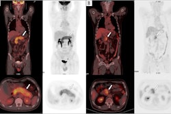
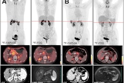
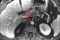
 Images of exendin PET/CT, DOTA-SSA-PET/CT, and CE-DWI-MRI (T1-weighted precontrast) of a study participant. Location of insulinoma in pancreas body is indicated with arrows. Only exendin PET/CT was true positive in this patient.Image available for republishing under Creative Commons license (CC BY 4.0 DEED, Attribution 4.0 International) and courtesy of the Journal of Nuclear Medicine.
In addition, in 13% of patients, a correct diagnosis was only reached after exendin PET/CT, the researchers noted.
“This study demonstrates the superiority of exendin PET/CT in a unique prospective comparison to all current routine imaging modalities for preoperative localization of benign insulinomas,” the group wrote.
Ultimately, the combination of exendin PET/CT and CECT provided the correct diagnosis in 53 of 54 participants in the study, the researchers noted.
“Exendin PET/CT combined with CECT could provide a one-stop-shop procedure for insulinoma localization, greatly reducing and simplifying the imaging work-up for the large majority of patients,” the researchers concluded.
The full study is available here.