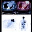
FDG-PET scanning could be better than CT at detecting local and distant metastases in patients with non-small-cell lung cancer, according to a study published in the July 27 New England Journal of Medicine.
Researchers from Groningen University Hospital in the Netherlands prospectively evaluated 102 non-small-cell lung cancer patients between 1996 and 1998 with CT and fluorodeoxyglucose (FDG) PET. They found that the latter modality was particularly effective for "distinguishing between intrapulmonary involvement and mediastinal lymph-node involvement," which can help determine if thoracotomy should be performed. (NEJM, July 27, 2000, Vol. 343:4, pp. 254-261).
"Indeed, with better staging of lung cancer, more patients will be unresectable and therefore unnecessary thoracotomies can be limited," said Dr. Harry Groen, one of the authors of the study.
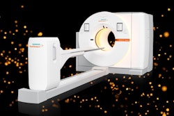
Patients were examined with a Siemens ECAT 951/31 PET scanner (Siemens/CTI, Knoxville, TN) that had a field of view of 10.8 cm and a resolution of 6 mm at full-width half-maximum. CT of the chest, upper abdomen, and adrenal glands was performed with a 120-kV, 124-mA Philips SR 7000 (Philips Medical Systems, Best, the Netherlands) with a slice thickness of 10 mm and a scanning time of one second per slice. In all but 13 patients undergoing CT, 200 ml of Omnipaque (Nycomed Amersham, Princeton, NJ) was administered. The imaging results were correlated with histopathological analysis.
In 29 of 32 patients, FDG-PET correctly detected mediastinal lymph-node metastases. FDG-PET also identified 28 out of 37 mediastinal lymph nodes as positive for metastatic tumors. Finally, FDG-PET correctly ruled out metastases in 60 out of 70 patients, according to the results.
"The sensitivity and specificity of PET for detecting mediastinal metastases were 91% and 86%, respectively," the authors wrote. "The overall negative predictive value was 95% and the positive predictive value was 74%."
FDG-PET produced a false-positive result in seven patients with reactive hyperplasia in the mediastinal lymph nodes, and false-negative results in one patient because of microscopic tumor residue. A third patient had false-positive results because of the scan’s inability to distinguish between paramediastinal primary tumor and mediastinal lymph nodes, according to the article.
Overall, FDG-PET correctly identified 11 distant metastases in 102 patients, with a sensitivity of 82% and a specificity of 93%.
In comparison, CT revealed enlarged mediastinal lymph nodes in 20 out of 37 patients who were positive for metastatic cancer on histopathology.
"CT correctly identified 46 of 70 patients who did not have mediastinal metastases on histopathological analysis and 24 of 32 patients who did have mediastinal metastases. The sensitivity of CT was 75% and the specificity was 66%," the report noted.
The FDG-PET results led to a change in cancer staging in 62 of 102 patients. The fact that non-small-cell lung cancer metastases "avidly incorporate [FDG] because they have an increased rate of glycolysis and overexpress the glucose transporter" made PET exams particularly adept at identifying tumors, they wrote.
At this stage, FDG-PET did have some drawbacks, including limited anatomical resolution, and the accumulation of FDG in the brain and urinary tract, making it difficult to determine if cancer had spread.
However, "the high negative predictive value of PET for mediastinal lymph-node metastases can be used to advantage…because invasive procedures are probably not necessary in patients with negative findings on PET in the mediastinum," the group concluded.
By Shalmali PalAuntMinnie.com staff writer
July 26, 2000
Related Reading
Lung cancer staging with PET accurately greenlights more resections than CT, June 20, 2000
CT and SPECT explored to better classify lung nodules, January 28, 2000
FDG-PET gains favor among referring doctors for cancer staging, treatment, June 7, 2000
Let AuntMinnie.com know what you think about this story.
Copyright © 2000 AuntMinnie.com


