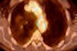
The ready availability of CT has resulted in the modality largely supplanting ventilation/perfusion (V/Q) scanning with gamma cameras to diagnose suspected pulmonary embolism, according to an upcoming study in the American Journal of Roentgenology.
By 2005, CT already was used six times more frequently than V/Q for pulmonary embolism (PE) diagnosis, according to the study, which examined several thousand hospitals across the U.S. CT's ready availability in major, small, and rural hospitals and its greater availability off-hours, along with recent clinical studies validating CT's utility for PE diagnosis, have contributed to the trend.
Study results will be published in the November issue of the American Journal of Roentgenology (Vol. 193:5, pp. 1324-1332). The lead author of the study was Dr. Mythreyi Bhargavan from the research department at the American College of Radiology (ACR) in Reston, VA. Bhargavan is also with the department of radiology and radiological science at Johns Hopkins University in Baltimore.
PE mortality
Pulmonary embolism is the third most common cause of cardiovascular death in the U.S. The study noted that major acute pulmonary embolism has a mortality rate of 26% when left untreated, with most deaths occurring in the first few hours after an embolism occurs. Because signs and symptoms of pulmonary embolism are nonspecific, a caregiver's ability to diagnosis the condition is limited without imaging.
Researchers used several sources for their survey:
- The Medicare Research Identifiable Files for 2005, which represent a random 5% sample of Medicare beneficiaries
- The Medicare provider of services file for 2005, which contains a list of hospitals serving Medicare patients and hospital data, such as size and medical school affiliation
- The American Hospital Association's AHA Guide 2006
In 2008, researchers also surveyed radiology departments of Pennsylvania hospitals about their use of CT and V/Q scanning for pulmonary embolism, service availability hours, and available equipment.
From those sources, patients were divided into two groups -- patients who received inpatient, outpatient, or emergency department service and were diagnosed with pulmonary embolism, and patients who were treated for symptoms that might have been due to pulmonary embolism. Those symptoms included chest pain, shortness of breath, syncope, and an actual diagnosis of pulmonary embolism.
CT over V/Q
Among the randomized Medicare dataset, the researchers found 3,270 hospitals with a total of 197,745 patients who presented with possible pulmonary embolism. Of these patients, 4,995 patients from 1,907 hospitals received a definitive diagnosis of PE.
Of the 4,995 patients, 33% were given a CT scan to diagnose their condition, while only 6% underwent V/Q scanning. The researchers found that CT was used roughly six times as often as V/Q, with V/Q exams making up 17% of the number of CT scans.
|
||||||||||||||||
| *Random 5% sample 2005 national Medicare data. Source: American Journal of Roentgenology. |
The researchers discovered that voluntary hospitals (35%) were more likely than for-profit (29%) or government hospitals (28%) to use CT to diagnose patients with pulmonary embolism symptoms. Private for-profit hospitals were more likely than other hospitals to use V/Q scanning (9%) than voluntary nonprofit hospitals (5%) and government hospitals (4%).
"We found that other factors being equal, large hospitals tend to use both CT and V/Q scanning more than other hospitals do," the authors wrote. "In other words, they evaluate patients in a generally more intensive style."
Local similarities
The researchers unearthed almost identical numbers in their review of Pennsylvania hospitals. An average of 39% of patients received a CT examination in the 98 Pennsylvania hospitals that had admitted at least one patient with a pulmonary embolism during the study period; 6% of patients with a pulmonary embolism diagnosis underwent V/Q scanning.
In 157 Pennsylvania hospitals with at least one patient with symptoms of pulmonary embolism, CT was used on an average of 3.5% patients, with an average of 0.5% for V/Q scanning.
As for the CT technology itself, 37 Pennsylvania radiology departments participated in the survey. There were an average of 2.7 CT scanners per department, with 16-slice to 64-slice systems accounting for one-third of those units. CT scanners with fewer than four detectors were in the minority, accounting for only 0.2 of the average 2.7 units.
The most common CT technology used for pulmonary embolism diagnosis was the 64-slice scanner. While 64-detector-row systems made up 20% of CT scanners in use, 40% of them were used to evaluate pulmonary embolism. Among those same facilities, 93% of them offered V/Q scanning, with 77% making it available 24 hours a day, seven days a week.
Continuing trend
The researchers speculated that the preference for CT rather than V/Q to diagnose pulmonary embolism has increased since 2005, in part because of the Prospective Investigation of Pulmonary Embolism Diagnosis (PIOPED) II trial, which illustrated the value of CT in this clinical application.
"Both the 2005 Medicare data and the 2008 survey, however, show much the same six-to-one preponderance of CT over V/Q scanning," the authors wrote. "Perhaps by 2005, V/Q scanning already was being used minimally except in the evaluation of patients with contraindications to CT, such as those with renal impairment or contrast allergy. In other words, CT likely was already a fully diffused technology."
In addition, citing the Pennsylvania results, CT is available 24/7 in 97% of the hospitals, while V/Q scanning was available around the clock in 77% of the facilities.
"We do not know whether this difference in availability is a consequence of physicians seeing relatively little need for V/Q scanning, and perhaps other nuclear medicine studies, at off hours and hospitals thus not employing staff to provide it," the researchers added, "or whether the staffing decision comes first, and the low use of V/Q scanning is partly a result of limited availability."
As for limitations, the researchers noted that the Medicare data excluded patients younger than 65 years. However, because the Pennsylvania hospital survey findings, which included patients of all ages, were so similar to the Medicare results, the authors said that it is "unlikely that the pattern of evaluation of nonelderly patients is quantitatively different from that of elderly patients."
By Wayne Forrest
AuntMinnie.com staff writer
October 20, 2009
Related Reading
Pulmonary CTA finds more than pulmonary embolism in children, August 25, 2009
At CT, not all pulmonary embolism are bad pulmonary embolism, February 4, 2009
Dual-energy CT helps distinguish pulmonary embolism from other perfusion defects, December 2, 2008
Multislice CT plus D-dimer testing sufficient for excluding pulmonary embolism, April 21, 2008
Study reinforces V/Q, PIOPED II guidelines for PE during pregnancy, February 15, 2008
Copyright © 2009 AuntMinnie.com


