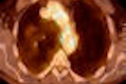
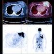
Researchers from Stanford University in Stanford, CA, will present a study that concludes that F-18 fluoride ion PET/CT may be helpful in predicting which metastatic lesions will be painful in the thoracolumbar spine. By doing so, the authors say it will allow for more effective treatment planning and could become an alternative to bone scanning with radiopharmaceuticals based on technetium-99m, which is in short supply.
In the study of 16 patients, researchers found "significantly increased" F-18 fluoride uptake in painful metastatic lesions compared to nonpainful lesions of the thoracolumbar spinal canal in subjects describing low back pain. The same patients also received an FDG-PET/CT scan.
Patients with spinal osseous metastases to the thoracolumbar spine and back pain had significantly higher mean F-18 fluoride ion uptake as measured by standardized uptake value (SUV) than patients with spinal metastases but no pain and those without metastatic disease. By comparison, no significant difference was observed in FDG uptake between the patients.
"Our study suggests that patients with osseous metastatic disease may benefit from F-18 fluoride ion PET/CT imaging," said co-author Dr. Sandip Biswal. "In our limited retrospective study, we found that F-18 fluoride ion PET/CT could predict which bone lesions were painful, while F-18 FDG-PET/CT could not."
He added that the results indicate the technique could be used in conjunction with FDG PET/CT. "F-18 fluoride ion PET/CT is essentially a bone scan 'on steroids,' and, therefore, cannot be used to detect or characterize extra-skeletal soft-tissue tumors," he added.
Biswal concluded that F-18 fluoride ion scans could replace bone scans with technetium-99m methylene diphosphonate (MDP), "but availability and cost will unfortunately slow its general acceptance in the medical community."


