The use of maximum standardized uptake values (SUVmax) from FDG-PET/CT scans may be a reliable, objective method for quantifying the activity and severity of Crohn's disease, according to a study in the July issue of the Journal of Nuclear Medicine.
Researchers from Rabin Medical Center in Petach-Tikva, Israel, based that conclusion, in part, on finding SUVmax to be significantly higher in abnormal segments of the ileum and colon than in normal segments of patients with known or suspected Crohn's disease.
The study, led by David Groshar, PhD, from Rabin's department of nuclear medicine, combined FDG-PET and CT enterography in a single examination, and compared the level of FDG uptake measured by SUVmax with the CT enterography patterns of disease activity found in patients with Crohn's disease (JNM, July 1, 2010, Vol. 51:7, pp. 1009-1014).
As Groshar and colleagues noted, CT enterography can detect enteric inflammation and extraenteric complications, as well as patterns of mural thickness, mural enhancement, mural stratification, increased attenuation of perienteric fat, and pericolic or perienteric hypervascularity, which indicate the presence of Crohn's disease.
FDG-PET/CT benefits
Whole-body FDG-PET/CT provides metabolic and anatomic information, as FDG accumulates in malignant tissues and areas of infection and inflammation.
"By combining the morphologic and physiologic patterns obtained by CT enterography with the FDG metabolic activity obtained by PET," the authors wrote, "PET/CT enterography as a single test may provide accurately fused morphologic, physiologic, and metabolic imaging that can be useful in the diagnosis, follow-up, and objective assessment of Crohn's disease."
The study included 28 patients suspected of having or known to have active Crohn's disease who were scheduled for CT enterography as part of a routine clinical workup. There were 11 males with a mean age of 42.1 years and 17 females with a mean age of 34.5 years. Patients who were younger than 18 years, pregnant, or unable to receive iodinated contrast material were excluded.
Patients fasted for a minimum of four hours before the intravenous injection of 185 MBq (5 mCi) of FDG. Patients also received bowel-distending negative contrast material for the CT enterography exam. All images were obtained from the midthorax through the pelvis with an eight-slice PET/CT scanner (Discovery ST, GE Healthcare, Chalfont St. Giles, U.K.).
Normal segments
In the analysis, the researchers noted 28 segments of normal-appearing ileum and 28 segments of normal-appearing colon. CT enterography wall thickness was 1.8 ± 0.6 mm, with no significant difference in the measured thickness between colon and ileum.
Mural enhancement among the 56 segments was 49.9 ± 14.7 HU, with enhancements, as expected, higher in the ileum than in the colon. PET SUVmax was 2.1 ± 0.69, with no significant difference in the measured SUVmax between colon and ileum.
In addition, the researchers detected 85 abnormal segments in 22 patients, with 57 abnormal segments (67%) in the ileum and 28 abnormal segments (33%) in the colon. Six patients had no abnormal segments noted on CT enterography and no pathologic uptake of FDG.
The analysis determined that the mean wall thickness was significantly higher in abnormal segments than in normal segments (8.6 ± 2.5 mm), with no significant difference in wall thickness between segments of ileum and segments of colon.
 |
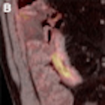
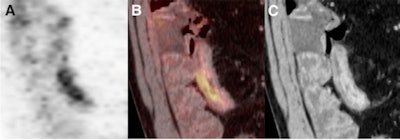
| The images depict intramural water attenuation on PET/CT enterography. Coronal PET image (A) shows increased uptake of FDG in distal ileum (SUVmax = 7). A fused PET/CT enterogram (B) shows FDG uptake in bowel wall. Coronal reformatted CT enterogram (C) shows abnormal segments with water attenuation by visual assessment in distal ileum (mural enhancement, 135 HU; wall thickness, 11 mm). Images courtesy of the Journal of Nuclear Medicine. |
Mean mural enhancement also was significantly higher in abnormal segments than in normal segments (113.4 ± 23.7 HU), with abnormal mural enhancement significantly higher in the ileum than in the colon (117.9 ± 21.7 HU and 104.2 ± 25.3 HU, respectively).
In addition, SUVmax was significantly higher in abnormal segments than in normal segments (5.0 ± 2.5), with no significant difference in SUVmax between segments of ileum and segments of colon.
'Good correlation'
The researchers also noted a "good correlation" between SUVmax measurements and wall thickness in the colon and ileum and no significant difference between the correlation in the ileum and the correlation in the colon.
A good correlation also was found between SUVmax and mural enhancement in the colon and ileum, with no significant difference between the correlation in the ileum and the correlation in the colon.
Based on those results, "SUVmax determination might be a reliable objective method for quantifying the activity and severity of Crohn's disease," the authors concluded. "Future studies are warranted to evaluate if combined PET/CT enterography has an important role in the clinical management of Crohn's disease."
By Wayne Forrest
AuntMinnie.com staff writer
July 9, 2010
Related Reading
CT-based Crohn's diagnosis takes aim at endoscopy, June 29, 2010
CT shows abdominal fat is unreliable Crohn's treatment target, April 14, 2010
MRI enterography shows shortcomings for Crohn's disease, February 23, 2010
ASIR cuts dose in Crohn's disease patients, December 4, 2009
FDG-PET/CT helps manage treatment for Crohn's disease, November 16, 2009
Copyright © 2010 AuntMinnie.com


