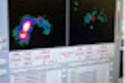
Gamma Medica is touting study results showing that molecular breast imaging (MBI) can detect invasive ductal carcinoma, ductal carcinoma in situ (DCIS), and invasive lobular carcinoma, and can play a beneficial role in evaluating extent of disease and presence of multifocal disease for surgical treatment planning.
The study, published in the April issue of the Journal of Nuclear Medicine, was conducted by researchers at the Mayo Clinic in Rochester, MN, to determine whether MBI is more sensitive than mammography in detecting additional foci of breast cancer in the ipsilateral and contralateral breasts, as well as in evaluating disease extent of biopsy-proven disease.
Patients with biopsy-proven breast cancer scheduled for surgery received a mammogram and MBI study prior to surgery. Patients with MBI studies showing additional sites of disease underwent additional diagnostic studies.
MBI studies were performed using Gamma Medica's LumaGem MBI system, which includes dual-head cadmium zinc telluride (CZT) digital detectors mounted on a modified mammography gantry. MBI patients were also injected with 296 MBq of technetium-99m sestamibi.
A total of 98 patients were enrolled in the study and received preoperative MBI and completed surgical resection. MBI detected additional disease greater than that identified by mammography and ultrasound combined, altering the surgical treatment in 12 (12%) of the 98 patients.
In seven (7%) of the 98 patients, MBI detected additional foci of cancer not seen on mammography. As a result, the patients' treatment plans changed from breast conservation to mastectomy. Final pathology confirmed the mastectomy decision.
In three (3%) of the 98 patients, MBI detected a significantly greater extent of disease than mammography that resulted in a change of surgical treatment plan from breast conservation to mastectomy.


